Colony Formation Assay Quantification

Tuft1 Promotes Osteosarcoma Cell Proliferation And Predicts Poor Prognosis In Osteosarcoma Patients In Open Life Sciences Volume 13 Issue 1 18

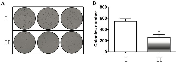
A B Representative Images And Quantification Of Colony Forming Assay Download Scientific Diagram
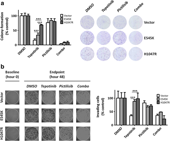
Pik3ca Hotspot Mutations Differentially Impact Responses To Met Targeting In Met Driven And Non Driven Preclinical Cancer Models Molecular Cancer Full Text
Inhibition Of Mir 155 A Therapeutic Target For Breast Cancer Prevented In Cancer Stem Cell Formation Ios Press
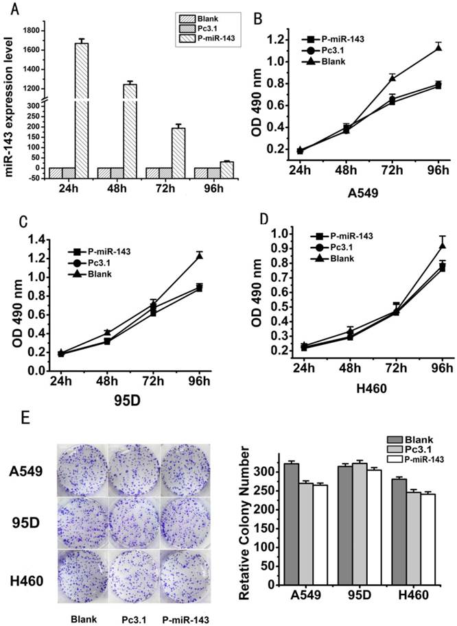
Microrna 143 Inhibits Migration And Invasion Of Human Non Small Cell Lung Cancer And Its Relative Mechanism

Tumor Spheroid Formation Assay Sigma Aldrich
Most measurements of attachment have used microscopy and colony counting, both of which are labor and time intensive To reduce the time and effort required to analyze bacteria attaching to plant tissues, we developed a quantitative realtime PCR (qPCR) assay to quantify attached A tumefaciens using the chvE gene as marker for the presence of the bacteria.
Colony formation assay quantification. ‘‘ColonyArea’’, which is optimized for rapid and quantitative analysis of focus formation assays conducted in 6 to 24well dishes ColonyArea processes image data of multiwell dishes, by separating, concentrically cropping and background correcting well images individually, before colony formation is quantitated. In particular, the colony formation assay has become a standard experiment for characterizing the tumor development in vitro However, quantification of the growth of cell colonies under a microscope is difficult because they are suspended in a threedimensional environment. Next, we investigated the effect of SUV39H1 silencing on EVderived miR744 mediated NSCLC cell proliferation using CCK8 assay and colony formation assay The results showed that the proliferation and colony formation in BEAS2BEVtreated A549 and H460 cells were retarded by siSUV39H11 treatment.
Cool plates for ~510 min at 4ºC to solidify agarose Do not stack plates Return plates to TC hood o Add 1 ml of growth media to each well Incubate plates at 37ºC and 5% CO2 for 10 to days,. This can be achieved using the colonyforming assay described here This protocol specifically applies to measurement of HeLa cells but can be used for most adherent cell lines with limited motility. The colony formation assay is an essential method for cancer research, enabling drug screens and radiation dosing to be conducted 15 The assay is performed by seeding cells at a low enough density such that individual cells can propagate to a sufficient colony area without impinging on a neighboring colony (Figure 1) 6, 7 At a set time point, adherent colonies are fixed then stained with Crystal Violet colorimetric dye, which allows for visual inspection of the culture vessel and.
Thus, the researcher must measure the diameter of each colony These techniques are necessarily subjective since the researcher has to manually trace the regions of interest in. Regulations for microbial threshold tests like TYM currently use nomenclature from culturebased methods to set acceptable thresholds It is necessary to convert the quantification cycle (Cq) value from the qPCR assay to colony forming units (CFU). Assay parameters can be easily adjusted to accommodate different metabolic states and strain backgrounds Highthroughput BioSpot counting technology with image recognition software adds additional power to this assay The BioSpot Analyzer counts up to 500 microscopic colonies per spot in a 96place format.
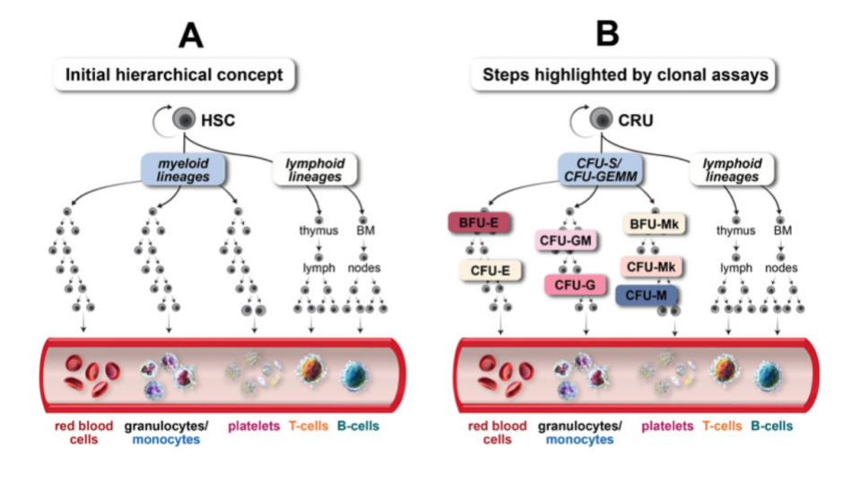
The colonyforming unit (CFU) assay is one of the most widely used assays for hematopoietic stem and progenitor cells (HSPCs) CFU assays allow measurement of the proliferation and differentiation ability of individual cells within a sample The potential of these cells is measured by the observation of the colonies (consisting of more differentiated cells) produced by each input progenitor cell. Subsequent counting of colony forming units (CFUs) In recent years a range of alternative, highthroughput (HT), methods relying on quantitative PCR (Neeley et al 05;. The primary method of monitoring anchorageindependent growth is the detection of soft agar colony formation, which measures proliferation by manual counting of colonies in semisolid culture media This traditional assay, though effective, is rather laborious in the initial setup and final quantification stages.
The primary method of monitoring anchorageindependent growth is the detection of soft agar colony formation, which measures proliferation by manual counting of colonies in semisolid culture media This traditional assay, though effective, is rather laborious in the initial setup and final quantification stages. It is necessary to convert the quantification cycle (Cq) value from the qPCR assay to colony forming units (CFU) State regulations for microbial thresholds currently use nomenclature from culturebased methods to set acceptable thresholds With this in mind, it was necessary for Medicinal Genomics to convert the quantification cycle (Cq) value from the PathoSEEK® qPCR Assay to colony forming units (CFU), which is reported when using culturebased methods. Cell colony as indicated A 50cell colony for Caco2 and HeLa cells corresponds to an area of 103x106 and 90x105 mm2, respectively These values were applied to color brightfield images of Crystal Violetstained colonies (C and F) Scale bar = 0 μm Automated Color Brightfield Imaging and Analysis of the Colony Formation Assay.
Lay et al 05) or genome probing with micro arrays (Bae et al 05) have gained popularity (Liu et al 04a). A colonyforming unit ( CFU, cfu, Cfu) is a unit used in microbiology to estimate the number of viable bacteria or fungal cells in a sample Viable is defined as the ability to multiply via binary fission under the controlled conditions Counting with colonyforming units requires culturing the microbes and counts only viable cells, in contrast with microscopic examination which counts all cells, living or dead. 004% DMSO) Average colony numbers were quantified and are graphed as mean (±SD) from triplicate wells (*** p Techniques Used Inhibition, In Vitro, In Vivo.
(C) Scans of representative MCF10A colony formation assays (D) Colony formation assay quantification of PMAtreated (25 ng/mL, 6 h) MCF10A cells relative to control (DMSO) Bars represent the average of 3 biological replicates ± SEM. I’m trying to perform the colony formation assay (or clonogenic assay) with a cancer cell line to study the effect of DNA repair inhibitors on the cytotoxicity of chemotherapeutic drugs. For the softagar assay, the formula commonly employed is A = πRr, where π = 314, and R and r are the longest and the shortest radii, respectively, of the colonies;.
Subsequent counting of colony forming units (CFUs) In recent years a range of alternative, highthroughput (HT), methods relying on quantitative PCR (Neeley et al 05;. One to two weeks are sufficient for HeLa cells to form colonies that are easy to count The time required to grow colonies from other cell types will vary based on the proliferation rates of each individual cell type Colonies of ∼ 50 cells are sufficient for counting by light microscopy at low resolution. Indeed, addition of exogenous VEGF to OP9 culture induces dispersion of the EC colony, rendering the quantification of the EC colony difficult 12 Nonetheless, the OP9 cell line provides a suitable condition for enumerating the number of clonogenic EC progenitors from both ES cell differentiation cultures and the actual embryos Because the proliferative capacity of EC may decrease on maturation, it should be noted that the proportion of EC progenitors clonable by this assay would decrease.
Vitro or in vivo assays 8;. Measurement of colonies made of fluorescently labeled cells is also an option;. In many assays biofilms are quantified by conventional culture plating method to get colony 10 forming units/count, which is an intensive procedure 1 Whereas other assays do use 96 well 11 microtiter plates for biofilm quantification as microtitre plate offers comparatively high 12.
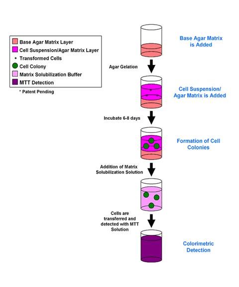
These data can also be used to calculate mean cells per colony Traditional methods for quantification of colonies by handcounting coupled with an assay for cell number (for example, DNA or mitochondrial) remains a viable method that can be used to calculate the mean number of cells per colony. Colony Formation Human bone marrow derived CD34 Hematopoietic Progenitor Cells were seeded at 3000 cells/well and cultured for 7 days in the presence or absence of growth factors/cytokines (hIL3, hIL6, hGCSF, hEPO) Colony quantitation was determined according to the assay protocol. The Soft Agar Colony Formation Assay allows testing of the therapeutic efficacy of compounds for anchorageindependent cell growth In the Soft Agar Assay, cells grow from single cells to cell colonies in an agar solution keeping them from the solid surface and allows growth in an anchorageindependent way.
The colony forming cell (CFC) assay, also referred to as the methylcellulose assay, is an in vitro assay used in the study of hematopoietic stem cells The assay is based on the ability of hematopoietic progenitors to proliferate and differentiate into colonies in a semisolid media in response to cytokine stimulation. True cellpercolony quantification, no areapercolony estimation needed;. Protein quantification using Bradford Assay with Coomassie Overview Protein binds to the Coomassie Plus reagent and causes a shift in signal from 465 to 595 nm with a simultaneous color change of the reagent from green/brown (or red/brown) to blue Pierce Coomassie Plus Protein Assay (Cat # ) has a detection range of 1µg/ml to 1µg/ml.
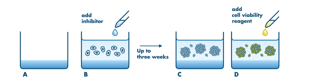
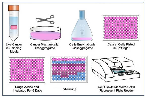
Soft Agar Colony Formation Assay Colony formation in soft or hard agar Last updated 8/12/14 By Madison Weg and Tim Starr Overview Use this protocol to test for cellular transformation exhibited by the ability to grow in an anchorageindependent setting Normal cells will not grow in soft agar due to anoikis, while transformed cells will. The Soft Agar Assay for Colony Formation is an anchorage independent growth assay in soft agar, which is considered the most stringent assay for detecting malignant transformation of cells For this assay, cells (pretreated with carcinogens or carcinogen inhibitors) are cultured with appropriate controls in soft agar medium for 2128 days. A cell viability reagent is added, and fluorescence is measured as an indirect quantification of colony growth in soft agar using Resazurin (D) Example Data The Aurora B inhibitor VX680 was tested for inhibition of the soft agar growth of A549 nonsmall cell lung cancer cells at indicated concentrations.
The bicinchoninic acid assay (BCA) is based on a simple colorimetric measurement and is the most common protein quantification assay BCA is similar to the Lowry or Bradford protein assays and was first made commercially available by Pierce, which is now owned by Thermo Fisher Scientific. One particular assay where livecell imaging can be useful is the colony formation assay This assay determines a cell’s ability to form a colony over a long period of time in culture 6 However, colony counting and assessment of colony sizes are primarily done manually, which can be a tedious process 6 To support researchers specific software developed for livecell imaging enables quantification of colony formation (colony size and colony count) over time, reducing manual labor. For the softagar assay, the formula commonly employed is A = πRr, where π = 314, and R and r are the longest and the shortest radii, respectively, of the colonies;.
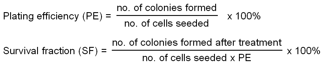
Clonogenic assay or colony formation assay is extensively used to measure in vitro cell survival based on the capacity of a single cell to grow into a colony When a cell divides and form a cluster of more than 50 cells, known as colony, is regarded as viable and traditionally counted manually 1 To obtain statistical accuracy such assays. Analyze the whole well continuously. Haarman and Knol 06), fluorescent labelling (Blasco et al 03;.
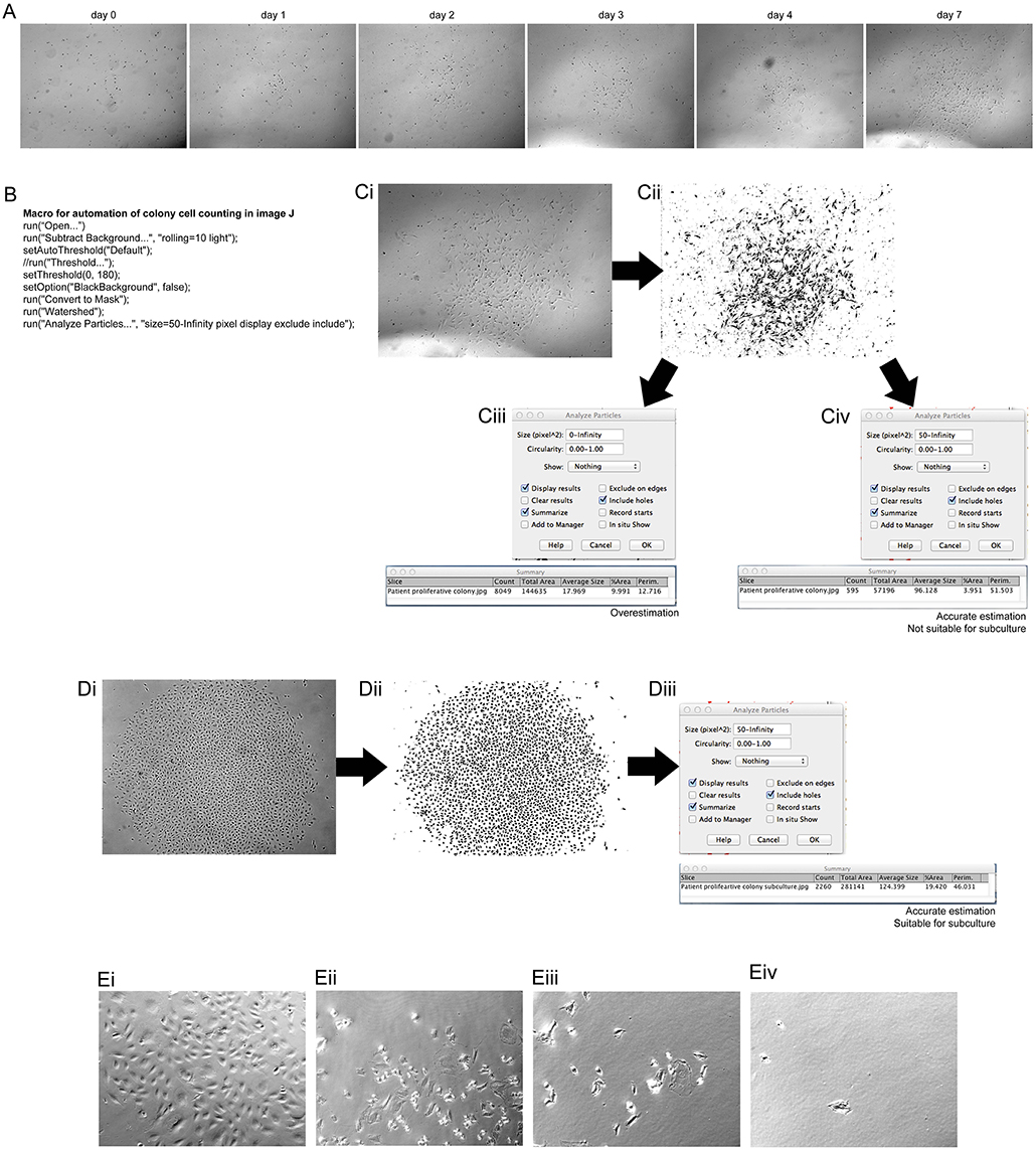
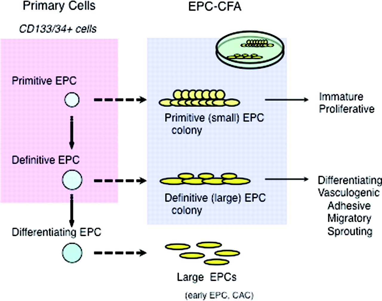
In cancer research, colony formation assay is a gold standard for the investigation of the development of early tumors and the effects of cytotoxic agents on tumors in vitro Quantification of cancer cell colonies suspended in hydrogel is currently achieved by manual counting under microscope It is challenging to microscopically quantify the colony number and size without subjective bias. Endothelial progenitor cells (EPCs) play a critical role in restoration of ischemic diseases However, the actual status of EPC development and the mechanisms of EPC dysfunctions in patients with various ischemic diseases remain unknown To investigate the detailed function of EPCs in experimental murine models, we have established an EPC colony forming assay (EPCCFA) in murine EPCs. The primary method of monitoring anchorageindependent growth is the detection of soft agar colony formation, which measures proliferation by manual counting of colonies in semisolid culture media This traditional assay, though effective, is rather laborious in the initial setup and final quantification stages.
Seventeen different L monocytogenes strains were tested using a modified crystal violet assay for their quantification The evaluation of their biofilmforming ability was carried out after a 48hour incubation period at 30ºC The media employed was the one proposed by Pan et al, 31 which was TSYEB with a supplement of glucose and sodium chloride. In particular, the colony formation assay has become a standard experiment for characterizing the tumor development in vitro However, quantification of the growth of cell colonies under a microscope is difficult because they are suspended in a threedimensional environment. One to two weeks are sufficient for HeLa cells to form colonies that are easy to count The time required to grow colonies from other cell types will vary based on the proliferation rates of each individual cell type Colonies of ∼ 50 cells are sufficient for counting by light microscopy at low resolution.
Final colonyforming units (CFUs) are calculated by correcting for the dilution factor 33 Viability Measured by FUN1 Staining Before heat ramp treatment ( see Note 7 ), 05 μl FUN1 cell stain and 5 μl Calcofluor White M2R are added to 1 ml yeast culture (OD 600 adjusted) with final concentrations of 5 and 25 μM, respectively. One particular assay where livecell imaging can be useful is the colony formation assay This assay determines a cell’s ability to form a colony over a long period of time in culture 6 However, colony counting and assessment of colony sizes are primarily done manually, which can be a tedious process 6. A Representative images of the colony formation assays using MCF7 or MCF7/TAMR cells under 0, 5, and 10 μM tamoxifen treatment for 14 days The bar graphs show the quantification of the colony formation assay data The data are presented as the mean ± SD of three independent experiments Student’s t test was used for statistical analysis.
The ‘gold standard’ for quantification of cytotoxicity is the colony forming assay This assay requires that colonies grow long enough to form visible colonies that are then counted manually We have created a new approach that enables miniaturization and automation. The initial cell number of each microorganism was determined by colony counting Each microorganism cell culture was diluted with medium and 190 μl of the cell culture was added to each well Then 10 μl of assay solution was added. Endothelial progenitor cells (EPCs) play a critical role in restoration of ischemic diseases However, the actual status of EPC development and the mechanisms of EPC dysfunctions in patients with various ischemic diseases remain unknown To investigate the detailed function of EPCs in experimental murine models, we have established an EPC colony forming assay (EPCCFA) in murine EPCs.
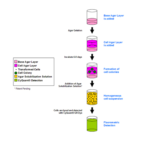
A clonogenic assay, also known as a colony formation assay is an in vitro cell survival assay It assesses the ability of single cells to survive and reproduce to form colonies 1 This assay was first described in the 1950s, where it was used to study the effects of radiation on cancer cell survival and growth and has subsequently played an essential role in radiobiology 2. Recently advances have been made in the soft agar colony formation assay to increase the speed and accuracy of quantitation These assays utilize a semisolid agar medium in which colonies are quantified using a fluorometric dye, thereby eliminating manual counting. MethoCult™ is the “Gold Standard” for the in vitro detection and quantification of hematopoietic progenitor cells in the colonyforming unit (CFU) or cell (CFC) assay, also known as the methylcellulose assay A wide range of MethoCult™ media formulations are available for CFU assays with hematopoietic cells from human and mouse tissues.
Lay et al 05) or genome probing with micro arrays (Bae et al 05) have gained popularity (Liu et al 04a). Quantify the ability of a single cell to form a colony A labelfree assayuse bright field imaging to identify the formation of a singlecell clone colony;. Haarman and Knol 06), fluorescent labelling (Blasco et al 03;.
Clonogenic assay or colony formation assay is an in vitro cell survival assay based on the ability of a single cell to grow into a colony The colony formation assay is a sensitive measure of compounds which requires cells to undergo several rounds of replication in order for the effect to be observed. In summary, our colony assay allows easy detection and quantification of functional progenitors within a heterogeneous population of cells In addition, the semisolid media format allows uniform presentation of extracellular matrix components and growth factors to cells, enabling progenitors to proliferate and differentiate in vitro. Recently advances have been made in the soft agar colony formation assay to increase the speed and accuracy of quantitation These assays utilize a semisolid agar medium in which colonies are quantified using a fluorometric dye, thereby eliminating manual counting.
Crystal Violet Cell Colony Staining 1L Fixing/Staining solution 05 g Crystal Violet (005% w/v) 27 ml 37% Formaldehyde (1%) 100 mL 10X PBS (1X) 10 mL Methanol (1%) 863 dH to 1L 1) Remove media (do not wash cells) 2) Add staining solution to cover dish 3) Stain for min at room temperature 4) Remove fix/stain solution and save. 321 Colonyforming unit assays CFU assays were first described in 1966 by Bradley and Metcalf as the shortterm quantitative assay for the measurement of differentiative potential of lineagerestricted progenitor cells (Bradley and Metcalf, 1966). Quantification of colony formation measuring absorption Colony formation was quantified following the method described by Kueng and coworkers In brief, the crystal violet staining of cells from each well was solubilized using 1 ml of 10% acetic acid and the absorbance (optical density) of the solution was measured on a Synergy H1 hybrid fluorescence platereader (BioTek, Winooski, VT, USA) at a wavelength of 590 nm.
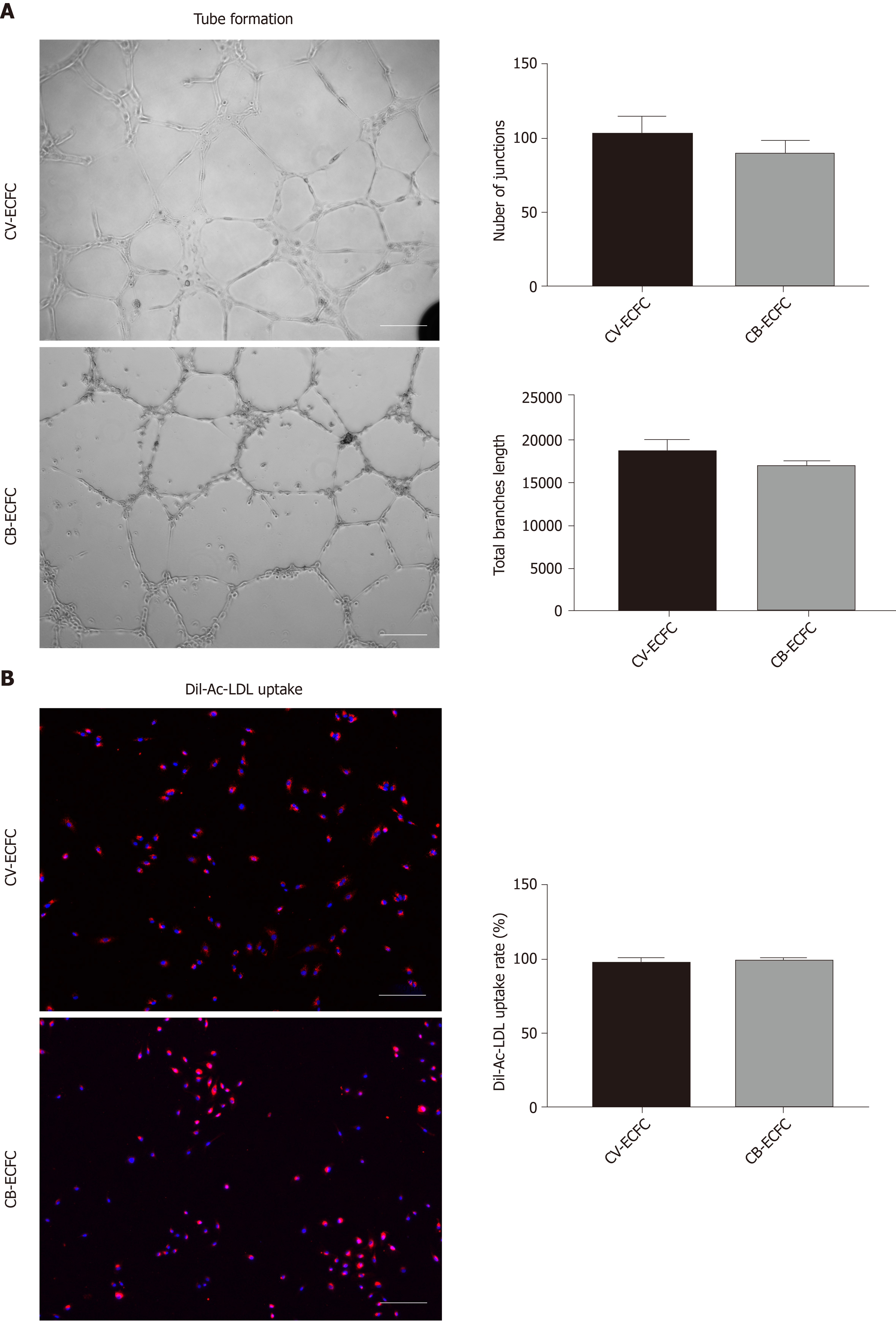
Protein quantification using Bradford Assay with Coomassie Overview Protein binds to the Coomassie Plus reagent and causes a shift in signal from 465 to 595 nm with a simultaneous color change of the reagent from green/brown (or red/brown) to blue Pierce Coomassie Plus Protein Assay (Cat # ) has a detection range of 1µg/ml to 1µg/ml. A colony assay that allows functional and quantitative assessments of single progenitor cells Numeration of the resulting colonies is used to calculate the frequency of the progenitor cells among the total plated cells The composition of cells within each colony is indicative of the lineage potential of the initiating progenitor. 9 In vitro Tube Formation Assay This assay involves plating endothelial cells onto a basementmembranelike substrate on which the cells form tubules within six to hours These tubules mostly contain a lumen, and the cells develop tight cellcell and cellmatrix contacts Quantification can be.
The ‘gold standard’ for quantification of cytotoxicity is the colony forming assay This assay requires that colonies grow long enough to form visible colonies that are then counted manually We have created a new approach that enables miniaturization and automation. The Soft Agar Assay for Colony Formation is an anchorage independent growth assay in soft agar, which is considered the most stringent assay for detecting malignant transformation of cells For this assay, cells (pretreated with carcinogens or carcinogen inhibitors) are cultured with appropriate controls in soft agar medium for 2128 days.
Plos One Colonyarea An Imagej Plugin To Automatically Quantify Colony Formation In Clonogenic Assays

Brd4 Is Dispensable In Naive Escs A Quantification Of The Colony Download Scientific Diagram
Www 2bscientific Com Getmedia 48cf7444 f3 4bbe B1ca Df2df3e965 Colony Formation Brochure 14 Pdf

Growth Of Epithelial Organoids In A Defined Hydrogel Broguiere 18 Advanced Materials Wiley Online Library

High Throughput Fluorescent Colony Formation Assay January 10

A Colony Formation Assay Representative Micrographs Left Panel And Download Scientific Diagram

Cytosmart Clonogenic Assay What Why And How

High Throughput Fluorescent Colony Formation Assay January 10
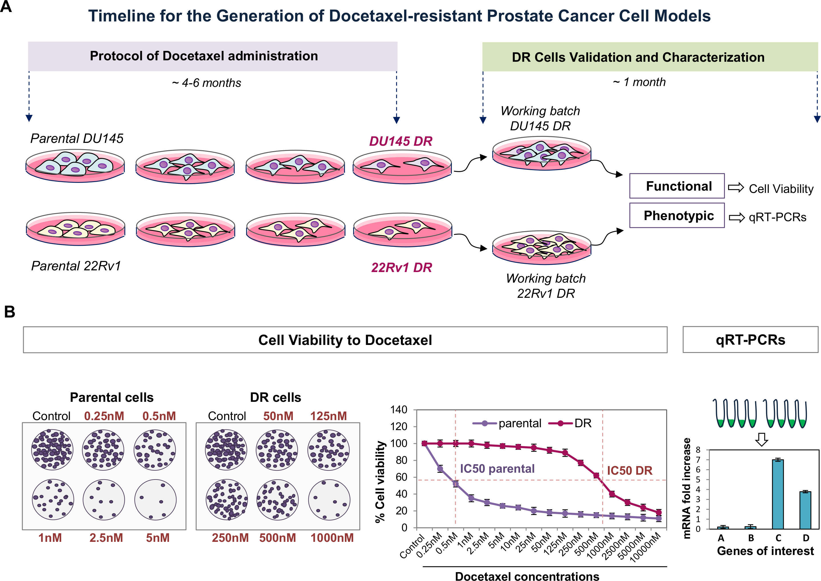
Generation Of Prostate Cancer Cell Models Of Resistance To The Anti Mitotic Agent Docetaxel Protocol

Colony Forming Assay Verification Of Ephrinb1 S Modulation Of Download Scientific Diagram
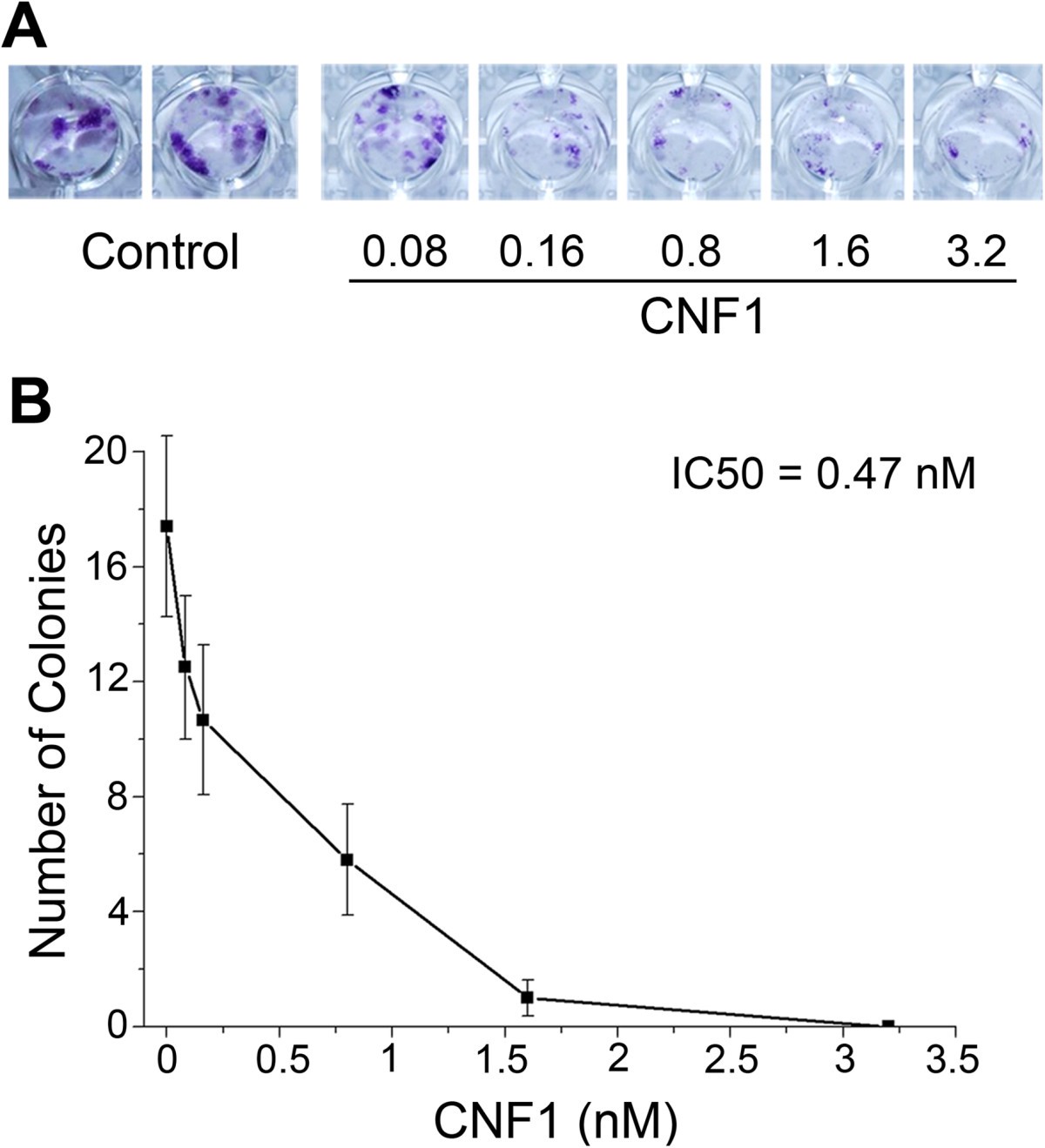
The Bacterial Protein Toxin Cytotoxic Necrotizing Factor 1 Cnf1 Provides Long Term Survival In A Murine Glioma Model Bmc Cancer Full Text

Biofilm Formation Assay In Pseudomonas Syringae

Mir 2b Silencing Activates Nf Kb And Promotes Aggressiveness In Breast Cancer Cancer Research

High Throughput Fluorescent Colony Formation Assay Metrolab Blog
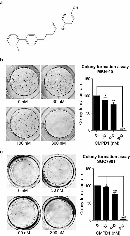
Cmpd1 Inhibited Human Gastric Cancer Cell Proliferation By Inducing Apoptosis And G2 M Cell Cycle Arrest Biological Research Full Text
Tumor Sensitivity Assay

Metformin Inhibits Tumorigenesis And Tumor Growth Of Breast Cancer Cells By Upregulating Mir 0c But Downregulating Akt2 Expression

Clonogenic Assay An Overview Sciencedirect Topics
Cytosmart Clonogenic Colony Formation Assay For Stem Cells
Plos One The Histone Demethylase Jarid1b Kdm5b Is A Novel Component Of The Rb Pathway And Associates With E2f Target Genes In Mefs During Senescence

A The Effects On Colony Formation Of Hepg2 Cells Treated With Pbs Download Scientific Diagram

High Throughput Fluorescent Colony Formation Assay January 10
Plos One Shrna Targeted Commd7 Suppresses Hepatocellular Carcinoma Growth

Figure 1 From Colonyarea An Imagej Plugin To Automatically Quantify Colony Formation In Clonogenic Assays Semantic Scholar
Plos One Colonyarea An Imagej Plugin To Automatically Quantify Colony Formation In Clonogenic Assays

Clonogenic Assay A B16 F10 B 058 C Jr8 Cells Were Treated Download Scientific Diagram

Figure 2 From Chloroform Fraction Of Serratulae Chinensis S Moore Suppresses Proliferation And Induces Apoptosis Via The Phosphatidylinositide 3 Kinase Akt Pathway In Human Gastric Cancer Cells Semantic Scholar
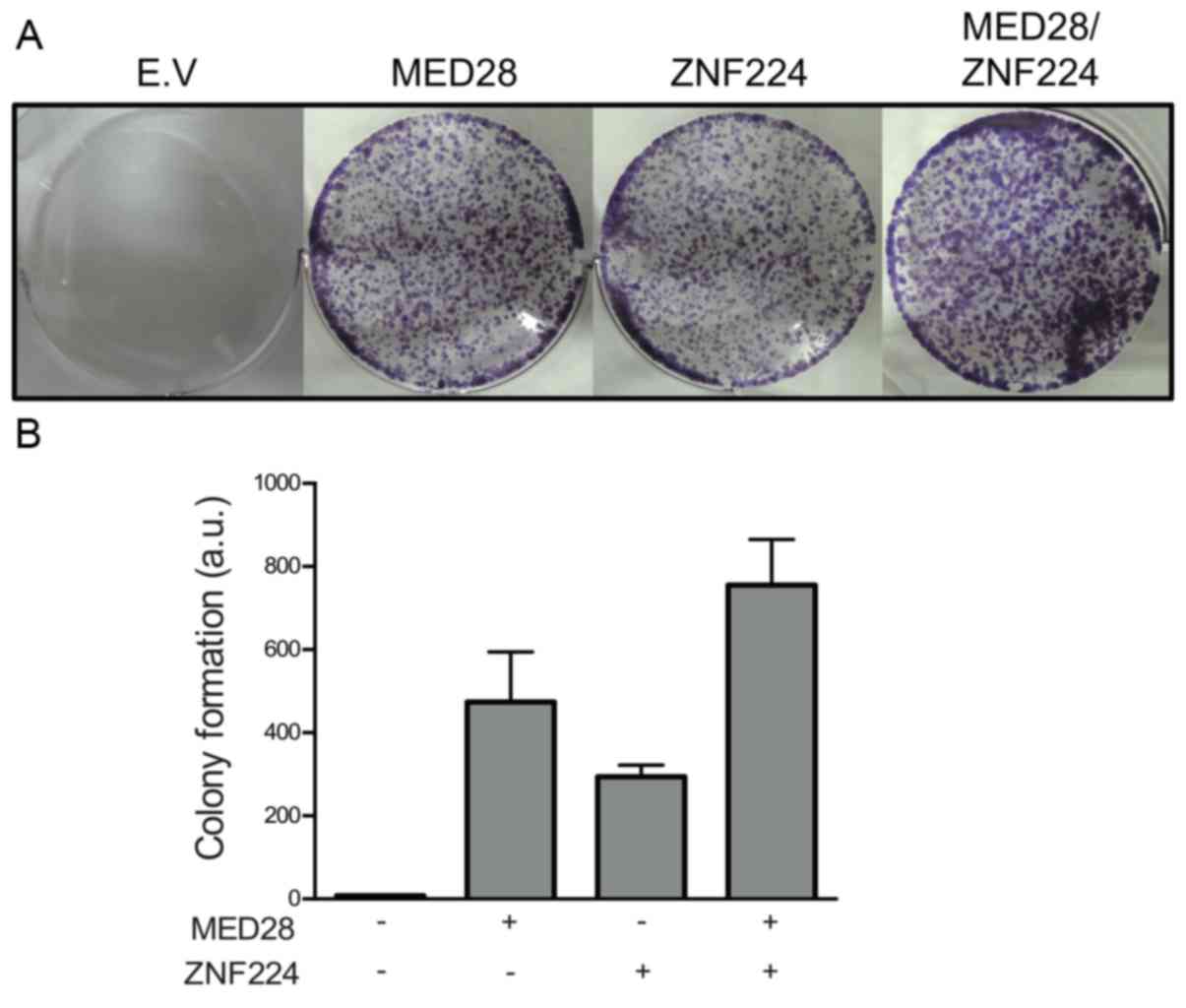
Med28 Increases The Colony Forming Ability Of Breast Cancer Cells By Stabilizing The Znf224 Protein Upon Dna Damage

Oxford Optronix Gelcount
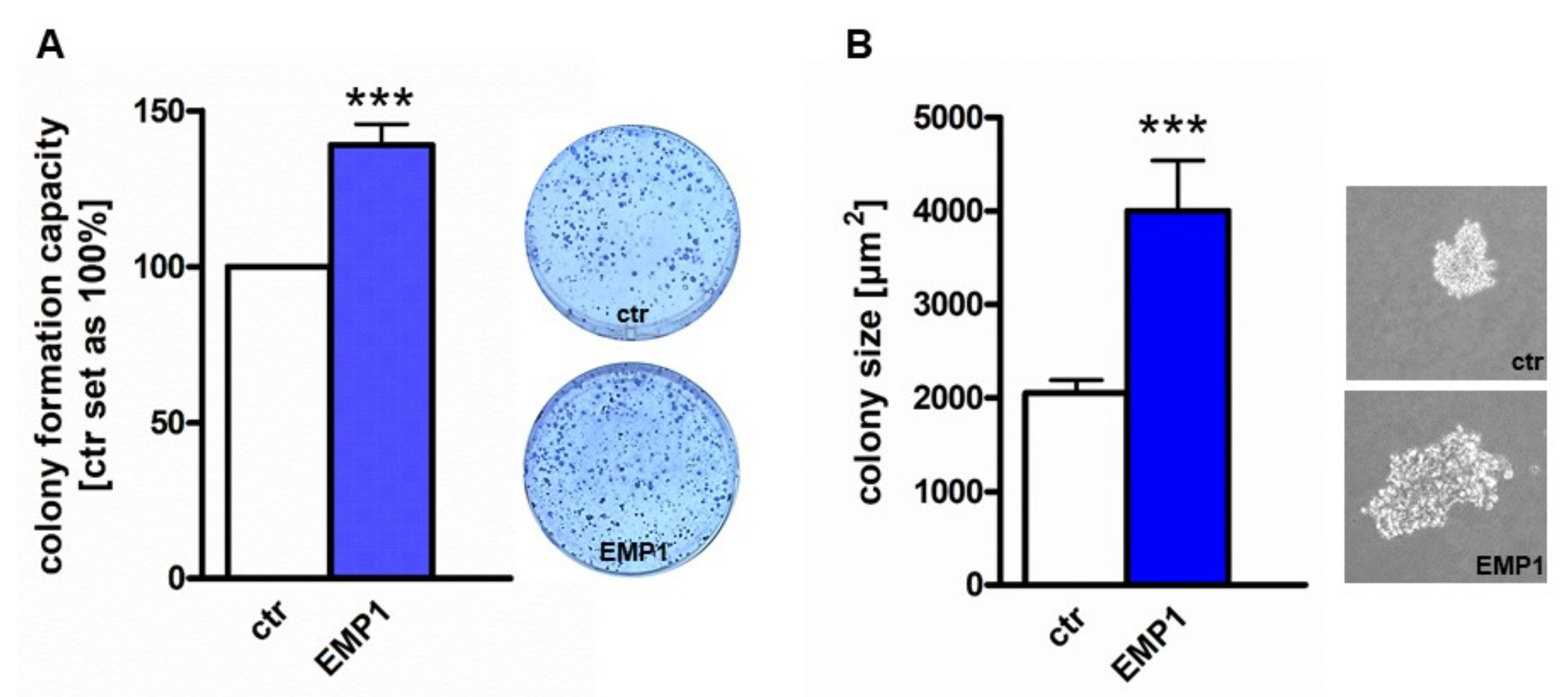
Ijms Free Full Text P53 Mir 34a And Emp1 Newly Identified Targets Of Tff3 Signaling In Y79 Retinoblastoma Cells Html

A Representative Colony Formation Assay B Quantitative Analysis Of Download Scientific Diagram

High Throughput Fluorescent Colony Formation Assay January 10

Pdf Colonyarea An Imagej Plugin To Automatically Quantify Colony Formation In Clonogenic Assays Semantic Scholar

Culture And Analysis Of Hematopoietic Progenitor Cells In Cfu Assays Stemcell Technologies
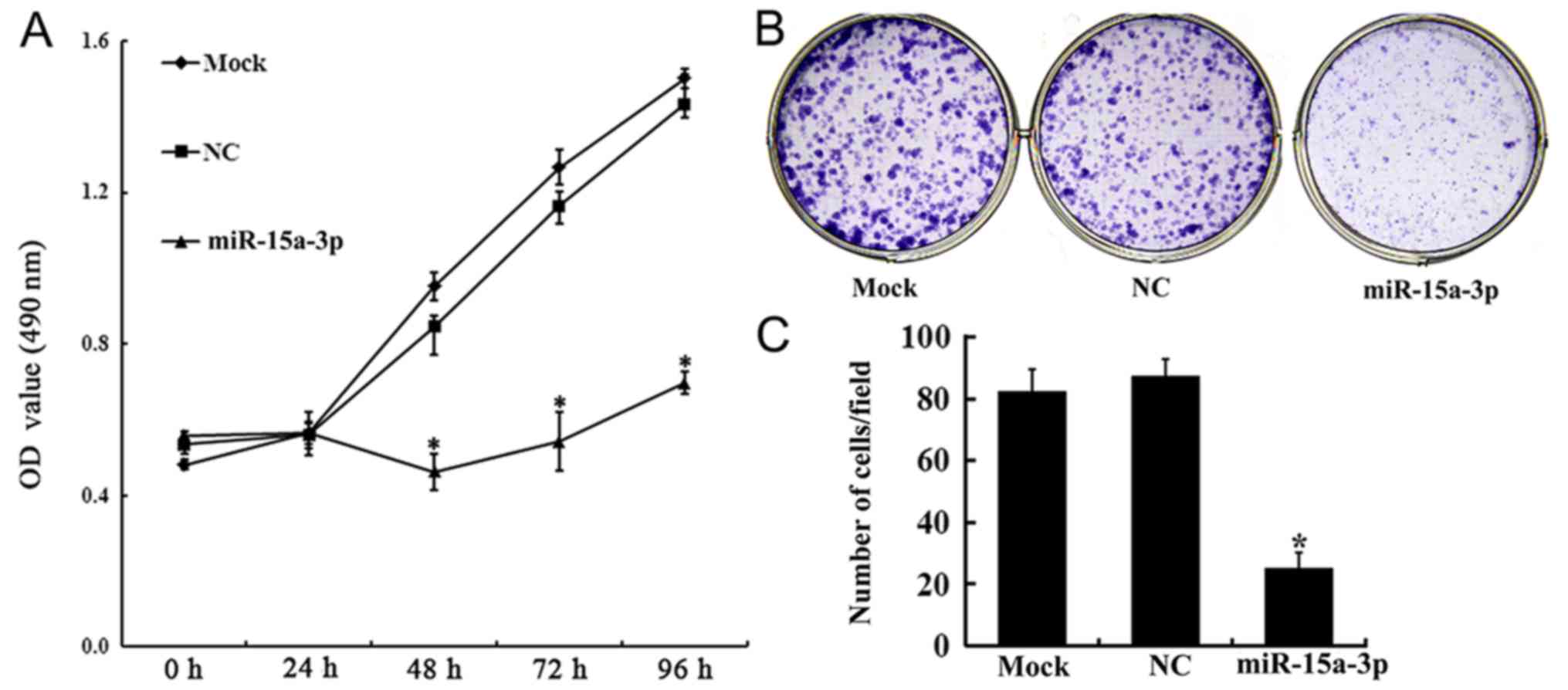
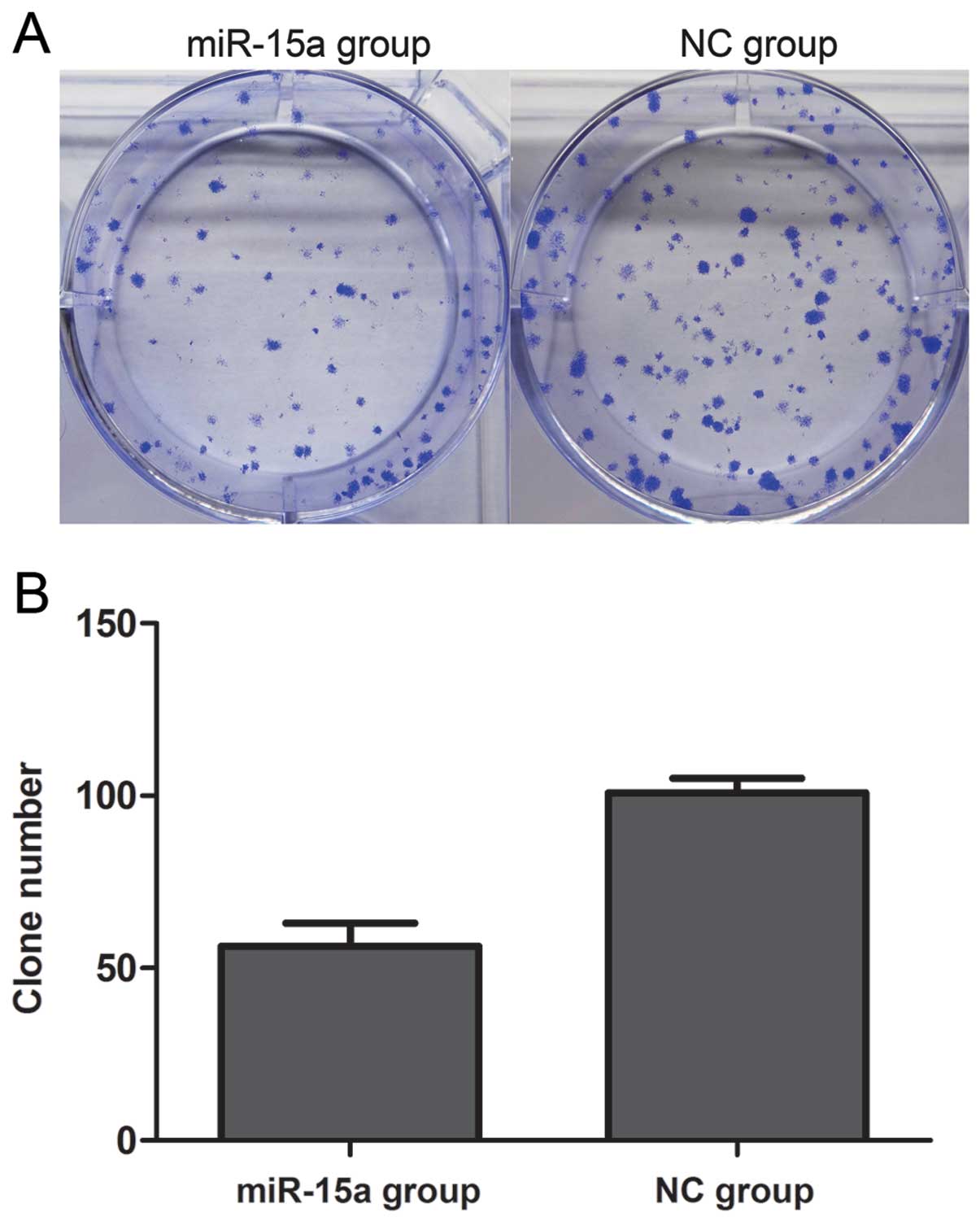
Mir 15a 3p Affects The Proliferation Migration And Apoptosis Of Lens Epithelial Cells

A Quantification Of Colonies Formed In Low Density Seeding Assay Download Scientific Diagram
Frontiers The Use Of Live Cell Imaging And Automated Image Analysis To Assist With Determining Optimal Parameters For Angiogenic Assay In Vitro Cell And Developmental Biology
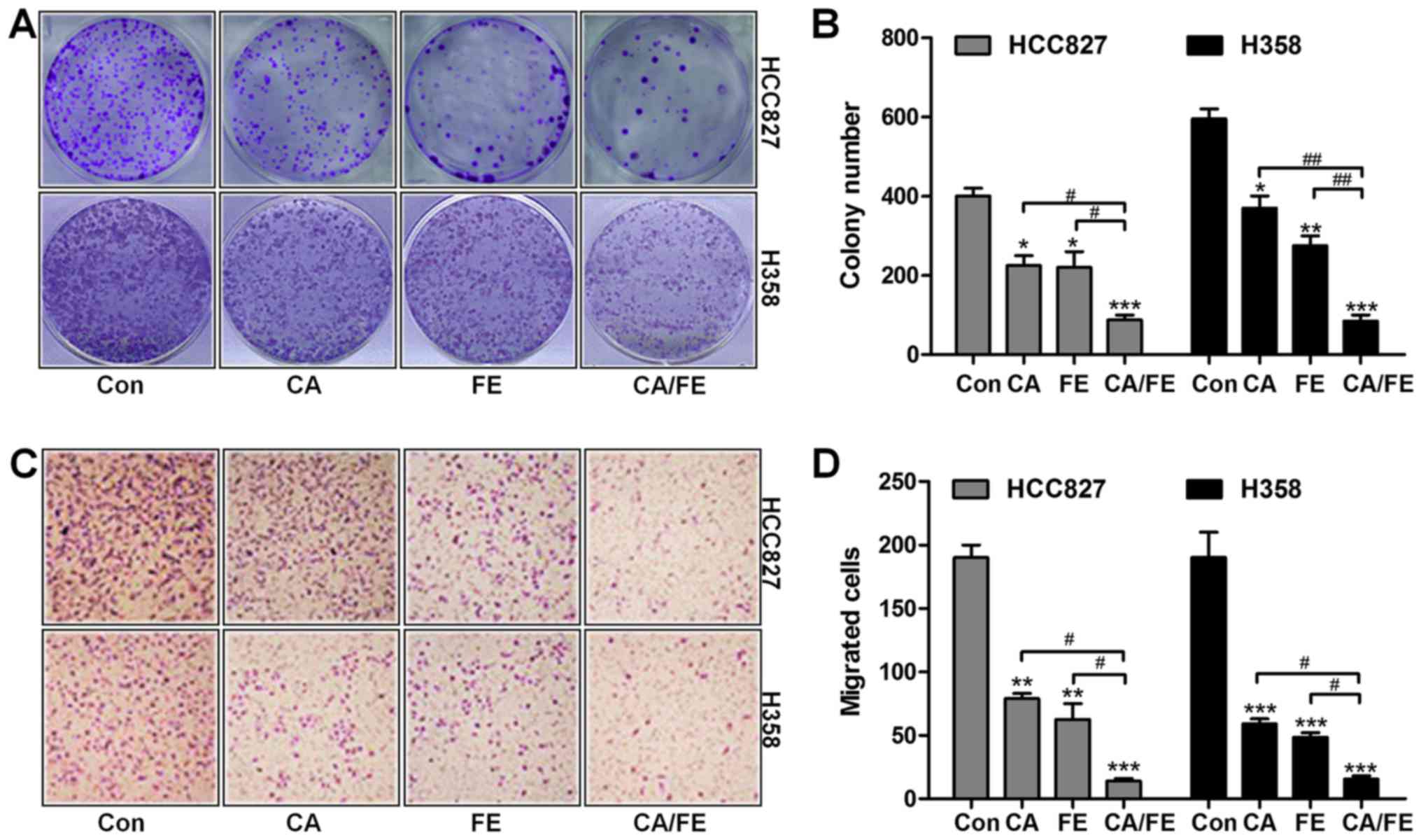
Carnosic Acid And Fisetin Combination Therapy Enhances Inhibition Of Lung Cancer Through Apoptosis Induction

An Hts Compatible 3d Colony Formation Assay To Identify Tumor Specific Chemotherapeutics Semantic Scholar
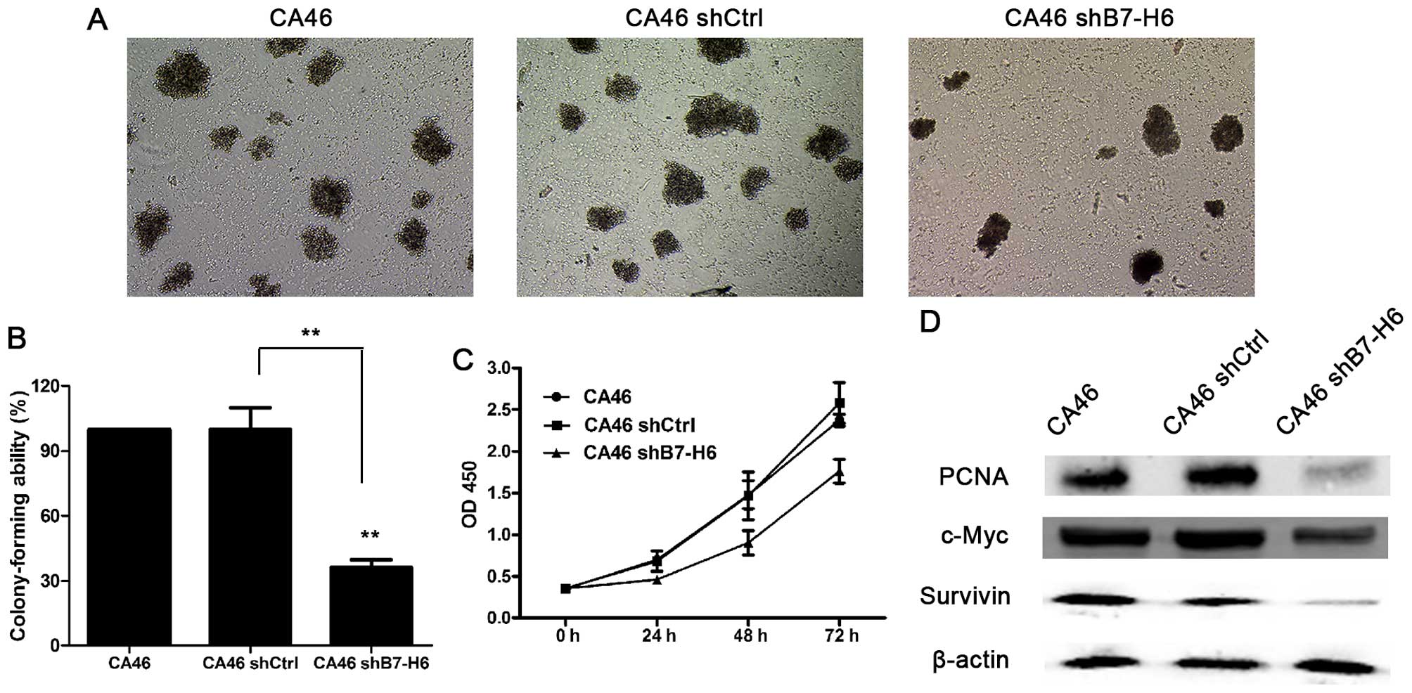
Knockdown Of H6 Inhibits Tumor Progression And Enhances Chemosensitivity In B Cell Non Hodgkin Lymphoma

Megacult Colony Assays Of Megakaryocyte Progenitors Cfu Mk
Www 2bscientific Com Getmedia 48cf7444 f3 4bbe B1ca Df2df3e965 Colony Formation Brochure 14 Pdf

Preclinical Assessment Of The Bioactivity Of The Anticancer Coumarin Ot48 By Spheroids Colony Formation Assays And Zebrafish Xenografts Protocol
Plos One Colonyarea An Imagej Plugin To Automatically Quantify Colony Formation In Clonogenic Assays

Imagej Macros For The User Friendly Analysis Of Soft Agar And Wound Healing Assays Biotechniques
Q Tbn And9gctmvp9fr7qyyqokcnx9gi48bab1whgbinevei1czx Lrcii0wqm Usqp Cau
Engelward Laboratory Microcolonychip Cell Survival Quantitation
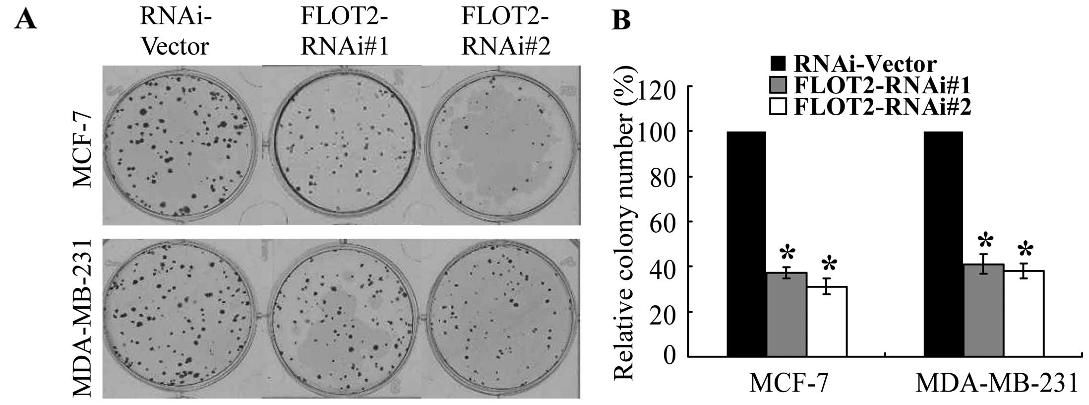
Knockdown Of Flotillin 2 Impairs The Proliferation Of Breast Cancer Cells Through Modulation Of Akt Foxo Signaling

Clonogenic Assay Youtube

Utilization Of The Soft Agar Colony Formation Assay To Identify Inhibitors Of Tumorigenicity In Breast Cancer Cells Protocol
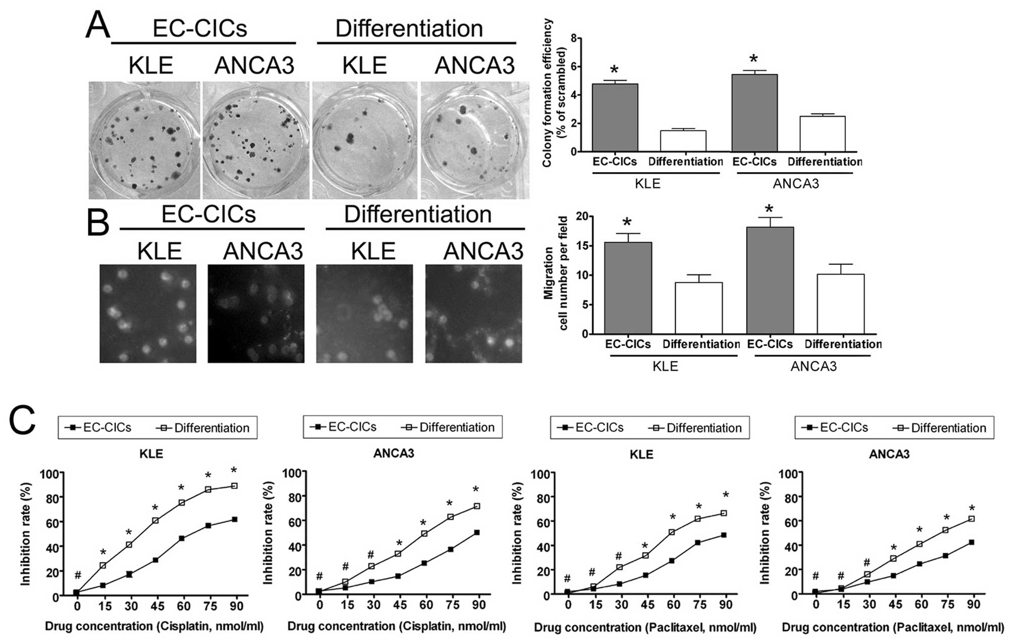
Isolation And Characterization Of Proliferative Migratory And Multidrug Resistant Endometrial Carcinoma Initiating Cells From Human Type Ii Endometrial Carcinoma Cell Lines

Cytosmart Clonogenic Colony Formation Assay For Stem Cells

High Throughput Fluorescent Colony Formation Assay Metrolab Blog

An Improved Crystal Violet Assay For Biofilm Quantification In 96 Well Microtitre Plate Biorxiv
Plos One Human Rad6 Promotes G1 S Transition And Cell Proliferation Through Upregulation Of Cyclin D1 Expression
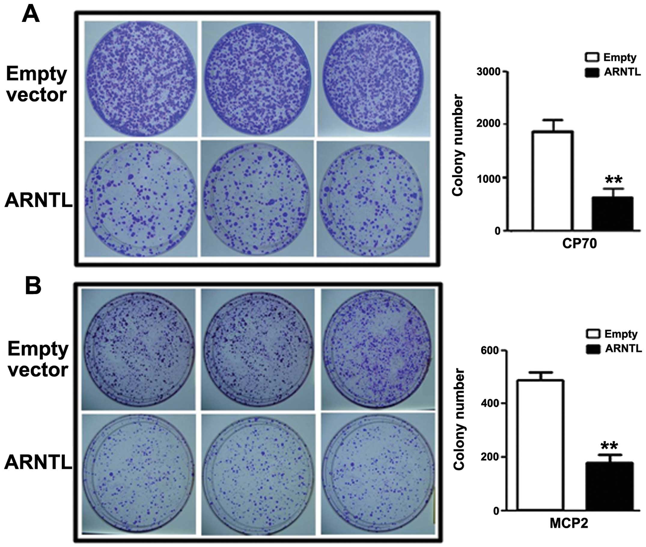
Epigenetic Silencing Of Arntl A Circadian Gene And Potential Tumor Suppressor In Ovarian Cancer

Clonogenic Assay Idea Bio Medical B

Oncogenic Transformation Of Human Mammary Epithelial Cells By Autocrine Human Growth Hormone Cancer Research
Www 2bscientific Com Getmedia 48cf7444 f3 4bbe B1ca Df2df3e965 Colony Formation Brochure 14 Pdf

Comparison Of The Colony Formation And Crystal Violet Cell Proliferation Assays To Determine Cellular Radiosensitivity In A Repair Deficient Mcf10a Cell Line Sciencedirect

Colony Formation And Cell Growth In Cwr22rv1 Cells Treated With Egcg Download Scientific Diagram

Utilization Of The Soft Agar Colony Formation Assay To Identify Inhibitors Of Tumorigenicity In Breast Cancer Cells Protocol
Q Tbn And9gcqj1jlgoj6etreht3kzoixepd Wx5pos9hqcwzu1ck Usqp Cau
Clonogenic Assay Creative Bioarray Creative Bioarray
Plos One The Architectural Chromatin Factor High Mobility Group A1 Enhances Dna Ligase Iv Activity Influencing Dna Repair

The Long Noncoding Rna H19 Promotes Tamoxifen Resistance In Breast Cancer Via Autophagy Journal Of Hematology Oncology Full Text

A The Fbxw7 Mrna Expression Level Of Hcc Cell Lines N 3 B Colony Download Scientific Diagram
Clonogenic Assay Creative Bioarray Creative Bioarray

Cytosmart Clonogenic Assay What Why And How
Www Cell Com Cell Reports Pdf S2211 1247 19 300 8 Pdf

Full Text Paeoniflorin Suppresses Pancreatic Cancer Cell Growth By Upregulating Dddt
Www Cell Com Cell Reports Pdfextended S2211 1247 19 300 8

High Throughput Fluorescent Colony Formation Assay January 10
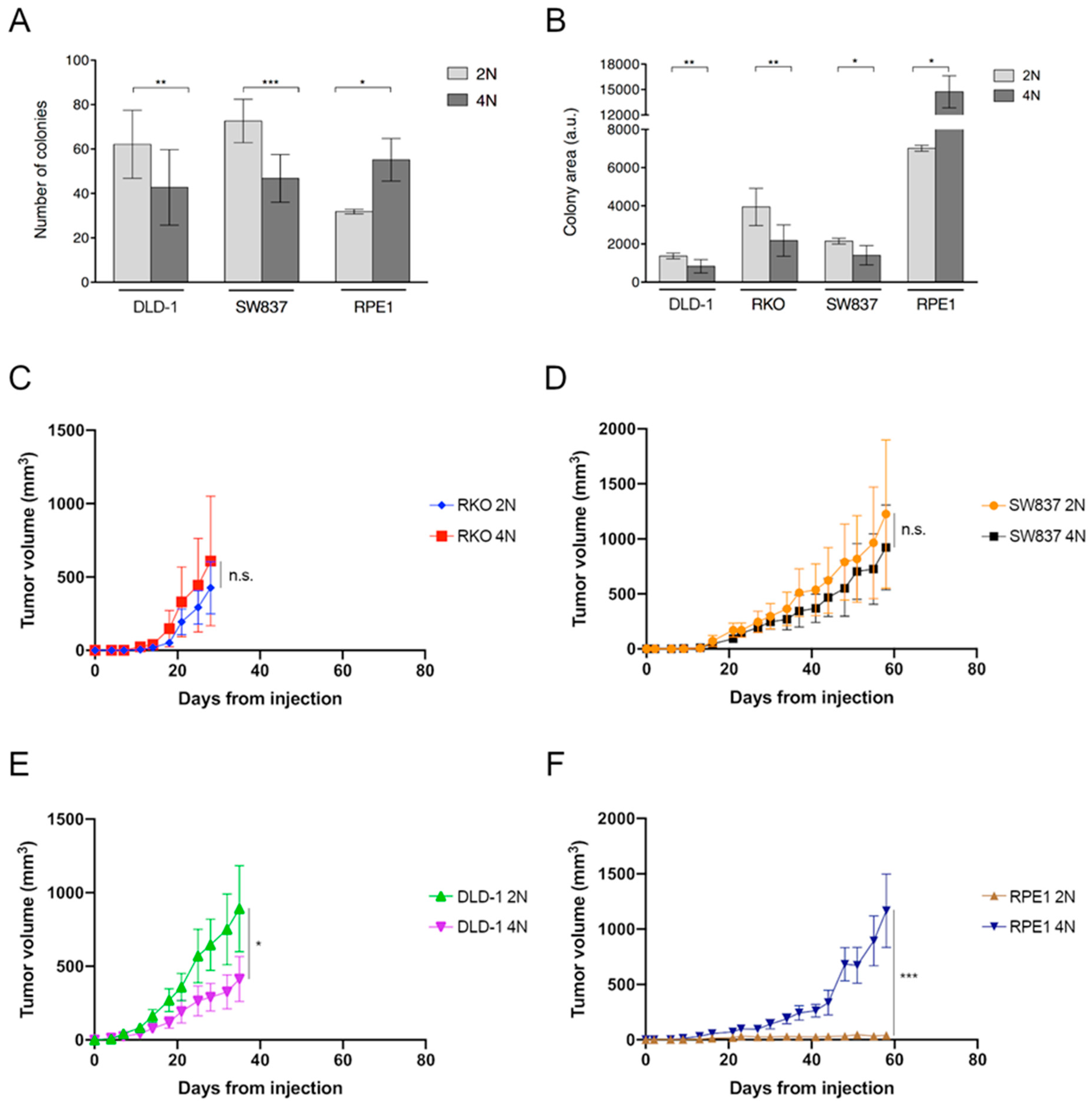
Cancers Free Full Text Tetraploidy Associated Genetic Heterogeneity Confers Chemo Radiotherapy Resistance To Colorectal Cancer Cells Html

Clonogenic Assay Of Nanoparticles Or Free Dox Or Elc In Hepg2 Cells Download Scientific Diagram

Cmpd1 Inhibited Human Gastric Cancer Cell Proliferation By Inducing Apoptosis And G2 M Cell Cycle Arrest
Cell Based Soft Agar Assays Reaction Biology
Clonal Isolation Of Endothelial Colony Forming Cells From Early Gestation Chorionic Villi Of Human Placenta For Fetal Tissue Regeneration

Idh1 R132h Mutation Increases Chemosensitivity To Tmz A Cell Colony Download Scientific Diagram
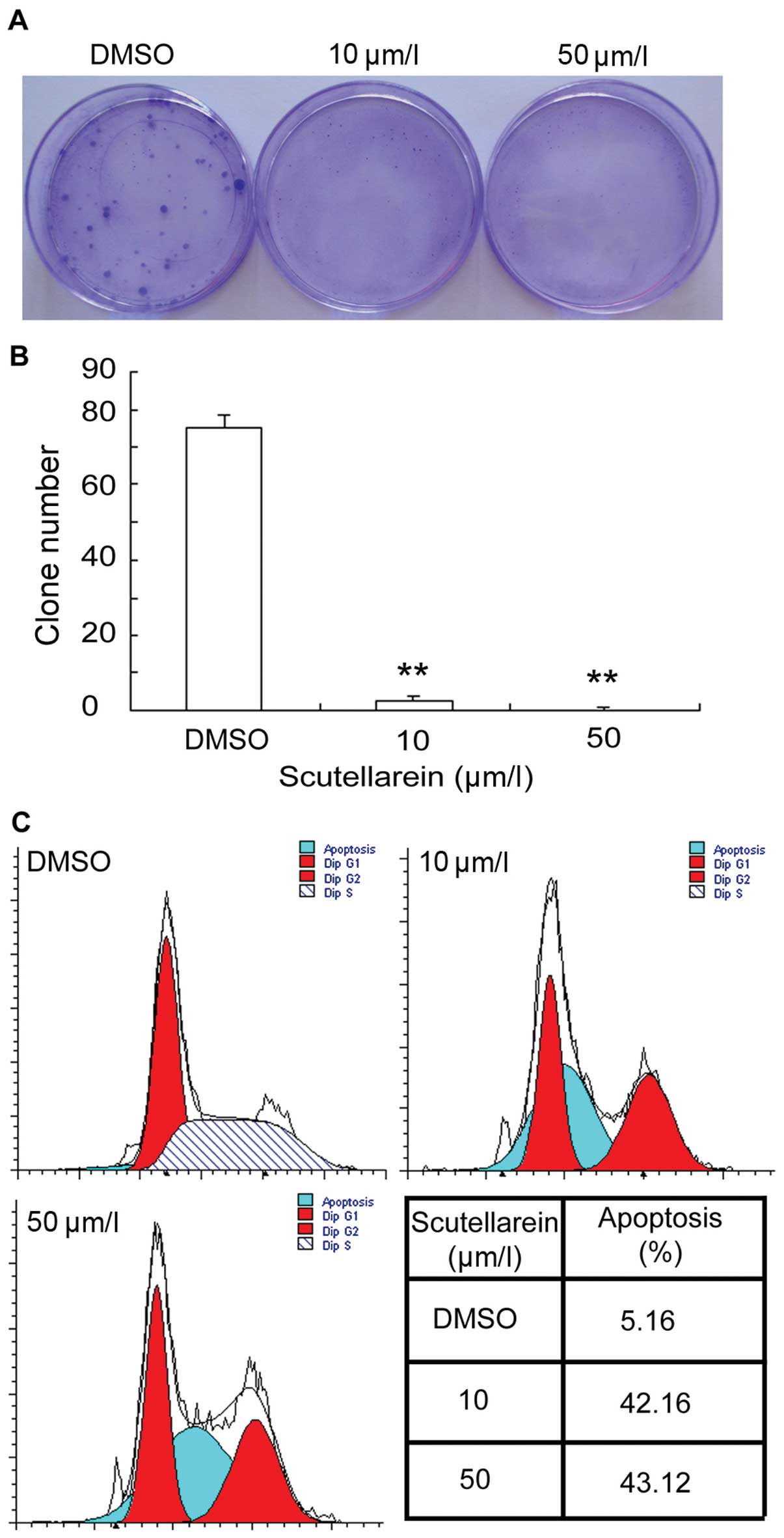
Scutellarein Inhibits Cancer Cell Metastasis In Vitro And Attenuates The Development Of Fibrosarcoma In Vivo
Q Tbn And9gcslemxvf V3xbbavhlwxx360golzqpnf4bfn Jgeeabr9mtoz9k Usqp Cau

Tumor Spheroid Formation Assay Sigma Aldrich
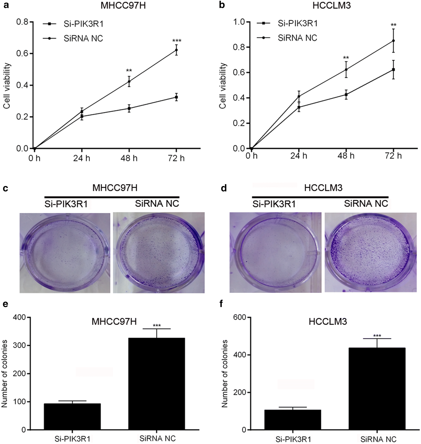
Overexpression Of Pik3r1 Promotes Hepatocellular Carcinoma Progression Biological Research Full Text

Cytosmart Clonogenic Assay What Why And How

Figure 2 From Small Interfering Rna Sirna Mediated Knockdown Of Notch1 Suppresses Tumor Growth And Enhances The Effect Of Il 2 Immunotherapy In Malignant Melanoma Semantic Scholar

Cytosmart Clonogenic Assay What Why And How

Cytosmart Clonogenic Assay What Why And How

Comparison Of The Colony Formation And Crystal Violet Cell Proliferation Assays To Determine Cellular Radiosensitivity In A Repair Deficient Mcf10a Cell Line Sciencedirect
96 Well Cell Transformation Assays Standard Soft Agar
Plos One Colonyarea An Imagej Plugin To Automatically Quantify Colony Formation In Clonogenic Assays
Cytosmart Clonogenic Colony Formation Assay For Stem Cells

Figure 2 S I Ammson I Is Required For Melanoma Growth And Survival
Soft Agar Colony Formation Assay Creative Bioarray Creative Bioarray
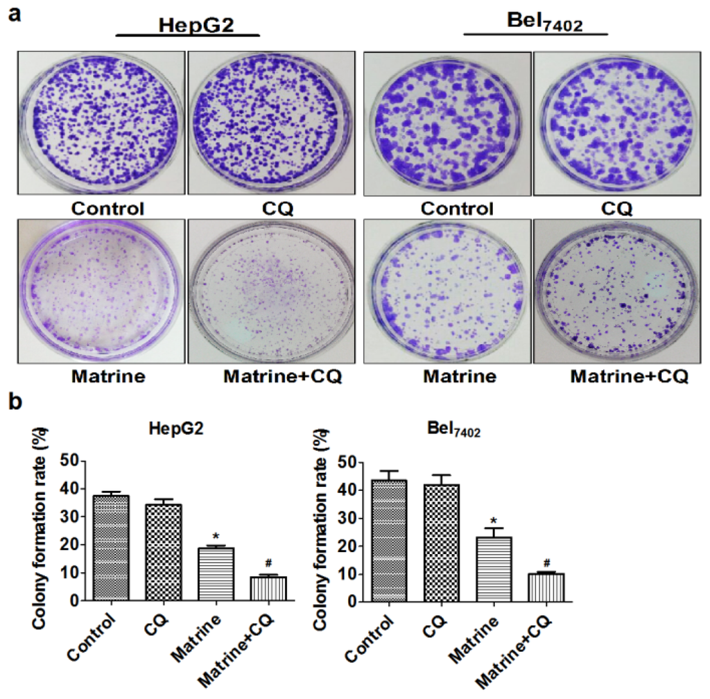
Ijms Free Full Text Blocking Autophagic Flux Enhances Matrine Induced Apoptosis In Human Hepatoma Cells Html
Mir 15a Is Underexpressed And Inhibits The Cell Cycle By Targeting Ccne1 In Breast Cancer

A Mts Assay B Quantification Of Colony Formation Assay P Download Scientific Diagram

The Soft Agar Colony Formation Assay Protocol


