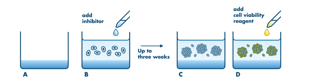
3d Colony Formation Assay
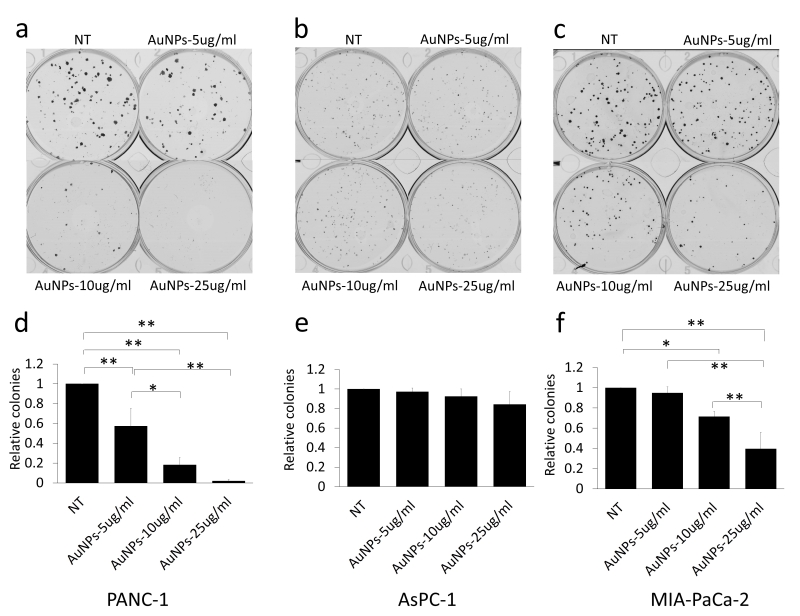
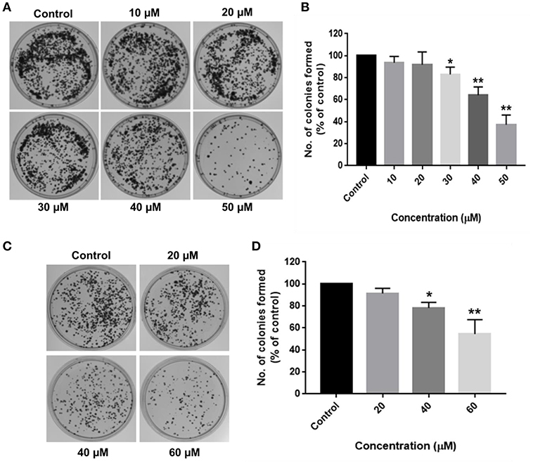
Gold Nanoparticles Sensitize Pancreatic Cancer Cells To Gemcitabine

Hematopoietic Colony Forming Unit Potential Assay Of Cd34 Cells Download Scientific Diagram
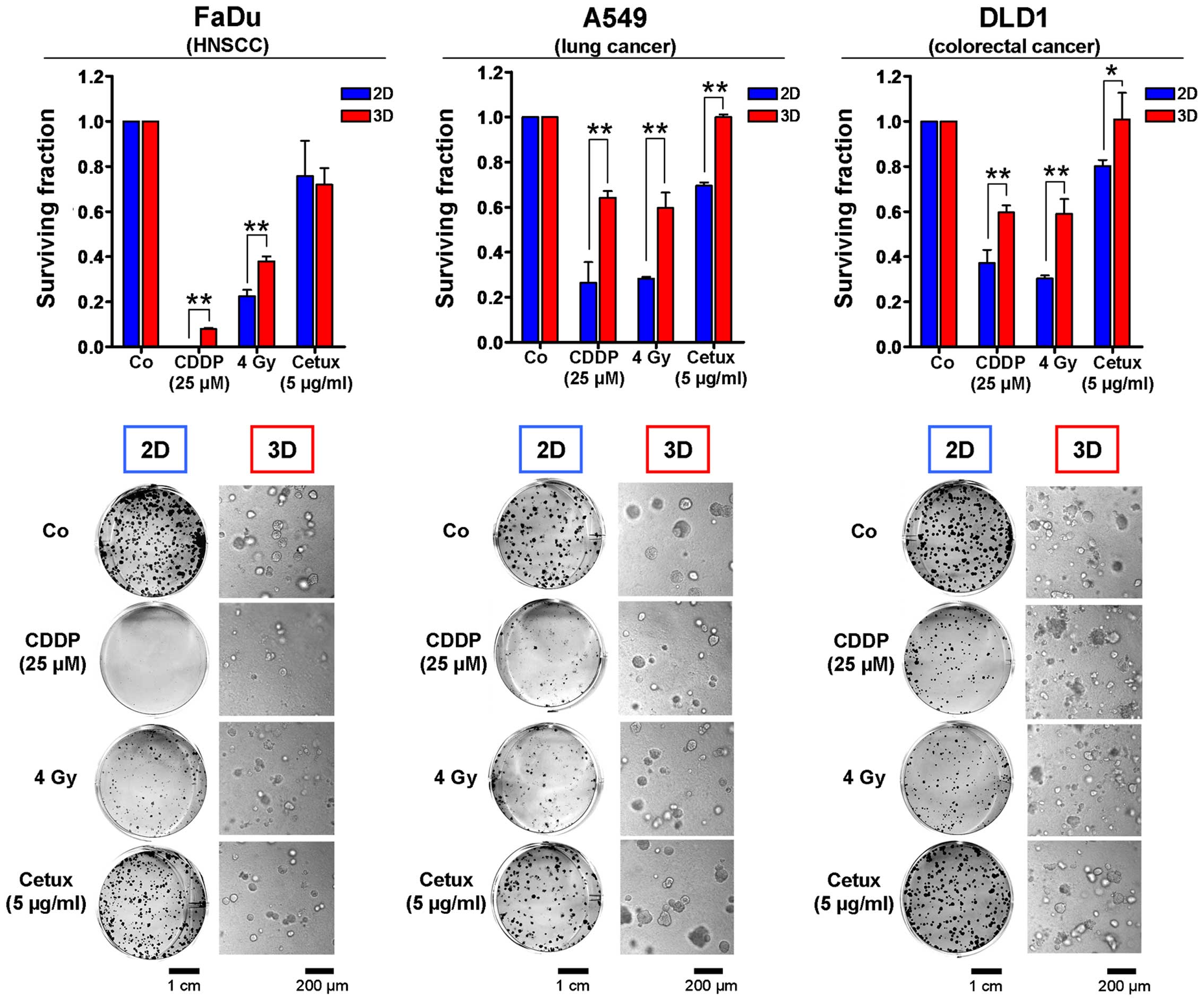
3d Matrix Based Cell Cultures Automated Analysis Of Tumor Cell Survival And Proliferation
Tumor Sensitivity Assay

Dark Colonies Are Formed From Dissociated Oneweek Old Murine Liver Or Download Scientific Diagram
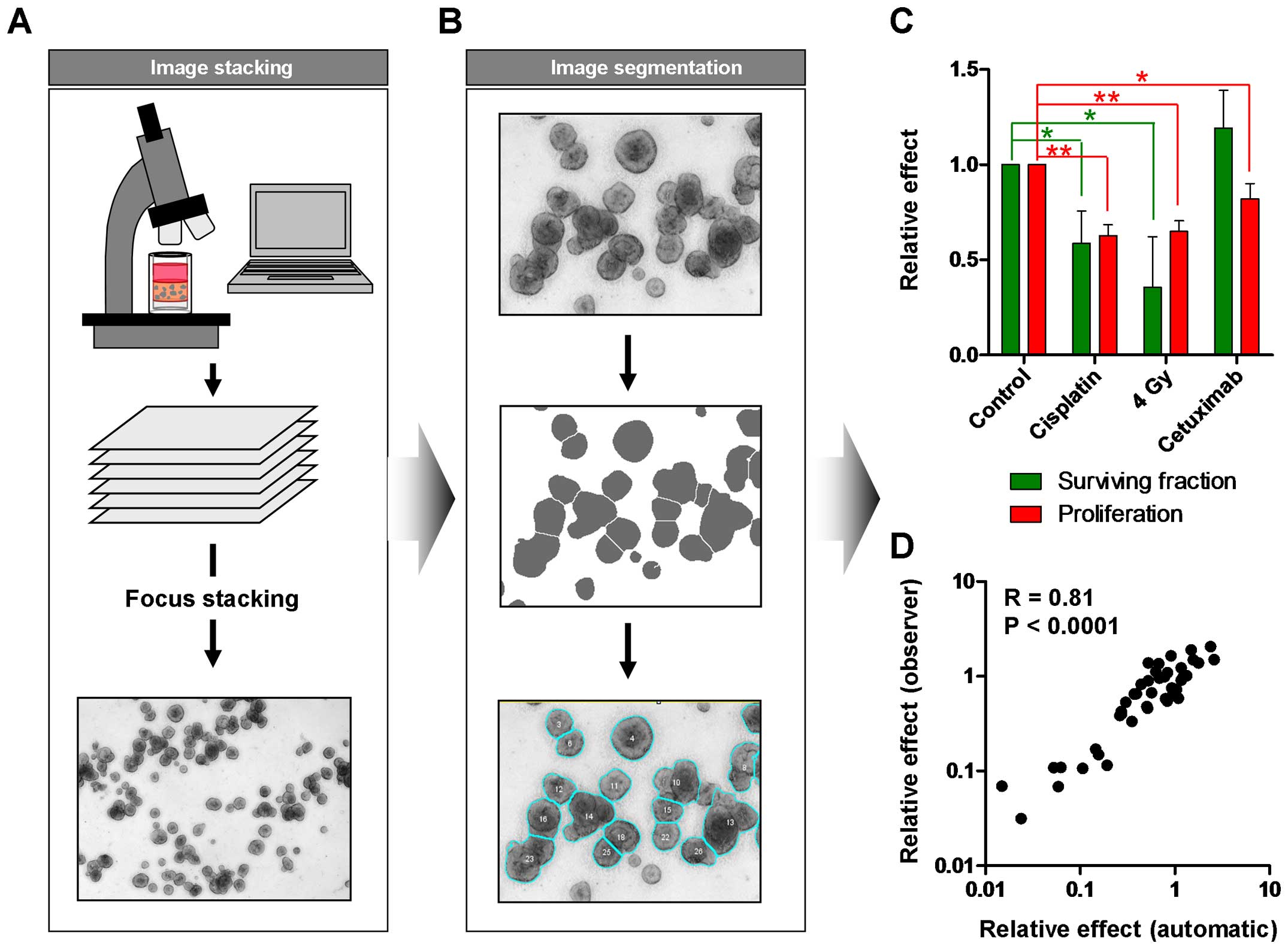
3d Matrix Based Cell Cultures Automated Analysis Of Tumor Cell Survival And Proliferation
This video protocol provides stepbystep instructions on how to consistently perform the Colony Forming Cell (CFC) Assay Tips are provided throughout the video to help optimize the assay procedure, including tricks to accurately evaluate and score colony formations.

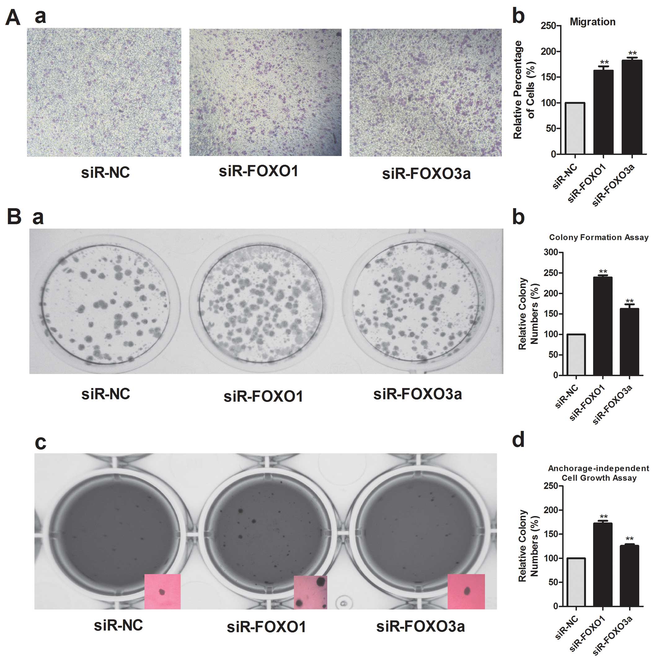
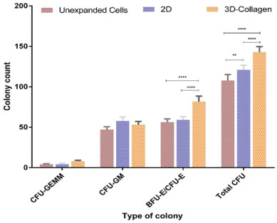
3d colony formation assay. The detailed colony formation assay processes was performed as described previously 25, 26 In short, different lentivirus infection GBC cell lines were seeded into 6well plates (about 800. Approximately 70 % of human CD34 hematopoietic cells accompanied with CD43 progenitor cells could be derived from this 3D induction system Colonyformingunit (CFU) assay showed that iPSCderived CD34 cells formed all types of hematopoietic colonies including CFUGEMM. To describe the responses of cell colonies, the most widely used quantification method is to count the number and size of cell colonies under microscope That makes the colony formation assay infeasible to be high throughput and automated.
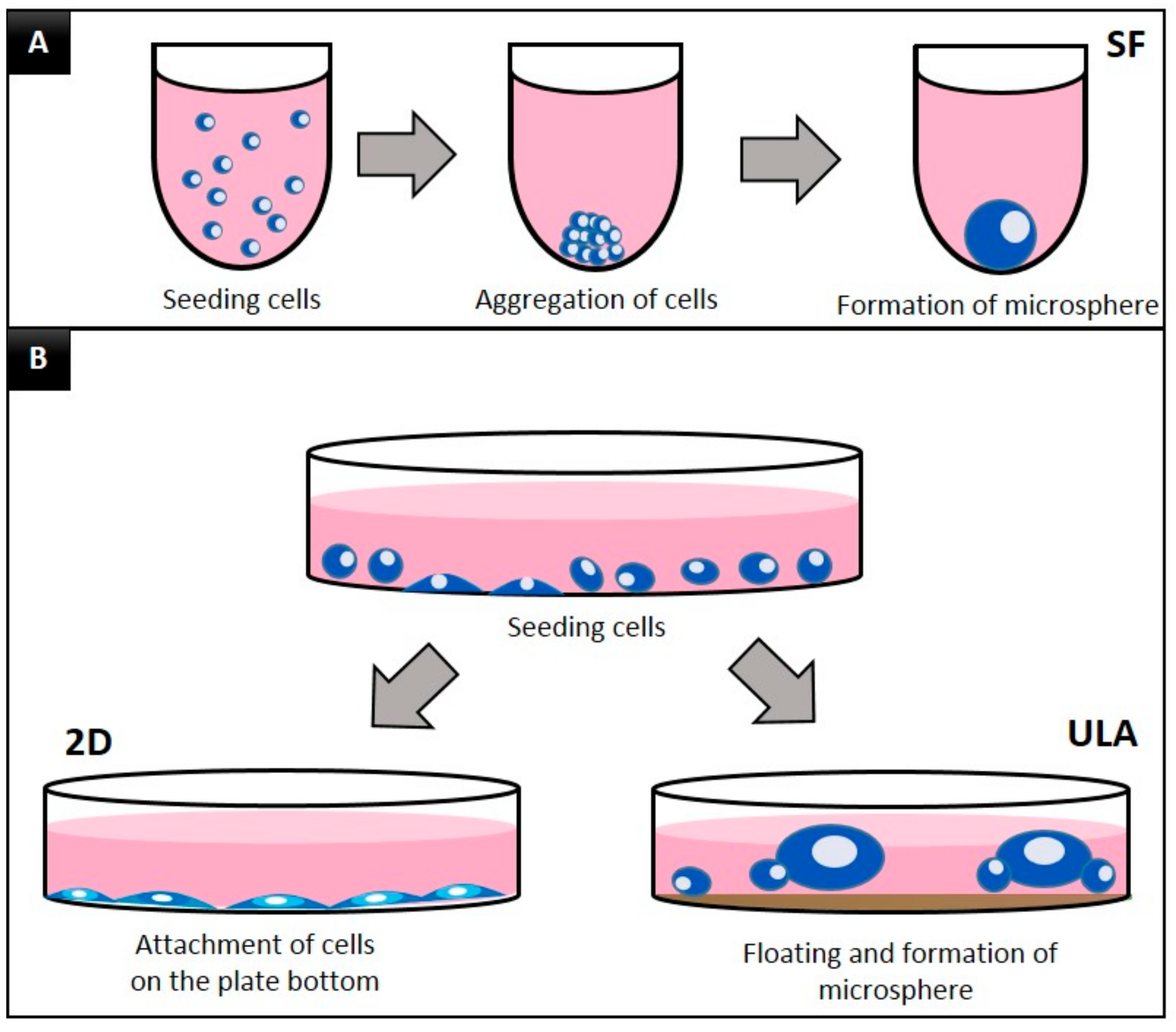
Colony formation assay is considered a 3D cell culture assay where cells grow independently of a substrate (also known as anchorageindependent growth) Only cancer cells can grow without a. 3D colony formation assay Asynchronously growing cells were trypsinized, counted and mixed with cell culture medium containing 05 mg/ml lrECM (cat no ;. Recipes Complete EpiCultB Medium (50 ml).
1) Matrigel is chilled on ice until thawed 46h (after C storage) 2) Cells are. BD Biosciences, Heidelberg, Germany) Then, 100 μl of this mixture was placed in 96well plates precoated with 50 μl of 1% agarose. 3D colony formation assay Clonogenic survival under 3D conditions was determined in a 3D colony formation assay as published previously Asynchronously and exponentially growing single cells were mixed with 05 mg/mL lrECM and placed in 96well plates LrECM was covered with cellculture medium.
In vitro Cytotoxicity Assay Colony Forming Assay (hamster cell line V79) In this assay the cytotoxicity of leachable substances released from the test item is assessed through the colony forming ability after treatment with various concentrations of the test item extract compared to the controls. Automated analysis of 3D colony formation assay can differentiate between effects on survival and proliferation (A) To account for limited field depth when imaging 3D cell cultures, multiple Zlevels are recorded and merged using an algorithm that preserves the sharpness of each plane (focus stacking). Formation methods, which are common in 3D culture, method versus the hanging drop and colony formation assays suggests that these three assays may be useful to test various cell properties.
A new approach to the colony forming assay 3D CoSeedis™ from abc biopply, a conical agarosebased microwell array, offers a new approach to the CFA The system was initially designed to open up 3dimensional (3D) organoid formation to a broader range of cells in cancer research To do so, the 3D CoSeedis™ matrix shows a unique and proprietary. In this study, we developed a 3D 384well agar colony formation assay using MB cells of molecular subgroup 3 that is associated with the highest level of metastasis Two fluorescence substrates, resazurin and glycylphenylalanylaminofluorocoumarin (GFAFC) that measure cell viability via distinct mechanisms were used to assess the growth of MB. The colony formation assay is an essential method for cancer research, enabling drug screens and radiation dosing to be conducted 15 The assay is performed by seeding cells at a low enough density such that individual cells can propagate to a sufficient colony area without impinging on a neighboring colony (Figure 1) 6, 7 At a set time.
It should be noted however that the application of 3D cell culture models to cancer drug discovery is not a new concept, indeed for almost 50 years, soft agar colony formation assays have been the gold standard in vitro method used to establish the transformation status of cells and for testing new drug candidates in low throughput , , , , , ,. Using the colony formation assay, we found that fibrin concentrations between 15 and 6 mg mL −1, which correspond to stiffness between 24 ± 10 Pa (SD, n = 6) and 270 ± 8 Pa (SD, n = 3), allowed efficient formation of round cysts (Figure 2B,F). Figure 2 3D mammary colonyforming cell assay Phasecontrast image of colonies derived from 3D culture (day 10) of mouse mammary single cells This image was obtained by using a Zeiss Observer Z1 microscope (5x/012 objective) and the AxioVision Rel 48 software Scale bar, 100 µm;.
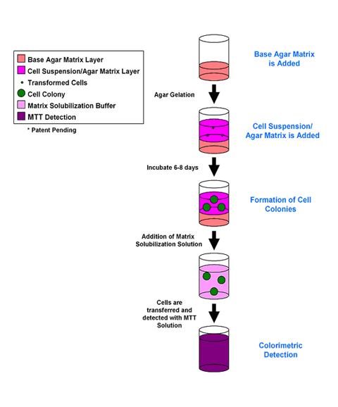
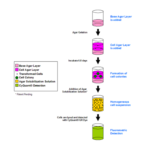
Colonies can be adherent or nonadherent (in semisolid 3D matrices such as soft agar or methylcellulose, or freefloating) GelCount thereby provides a powerful and costeffective alternative to the subjective and labourintensive task of manual colony counting in the gold standard colony forming cell assay, also referred to as the clonogenic assay, the cell survival assay or the tumor cloning assay. The in vitro spheroid formation assay includes methods to generate colonyforming units (CFUs) in both semisolid media and as 3D tumorsphere aggregates The tumorsphere formation efficiency (TFE assay) indicates the percentage of cells within a cancer cell culture that are capable of forming a sphere from a single cell. SOFT AGAR ASSAY FOR COLONY FORMATION Note All volumes are calculated to cater for four plates per point Base Agar 1 Melt 1% Agar (DNA grade) in microwave, cool to 40 °C in a water bath Warm 2X DMEM/F12 additives to 40 °C in water bath Allow at least 30 minutes for temperature to equilibrate.
Soft Agar Assay Protocol 1 Preparation of Base Agar a Dissolve 1% agarose (Difco Agar Noble) in sterile H, cool to 42°C in water bath Use autoclaved 125 mL screwtop bottle b Warm 2X DMEM w/% FBS and antibiotics to 42° in water bath i. An HTSCompatible 3D Colony Formation Assay to Identify TumorSpecific Chemotherapeutics Journal of Biomolecular Screening, Vol 18, No 10 Alternative Splicing of the RAGE Cytoplasmic Domain Regulates Cell Signaling and Function. 3D Matrigel colony assays what's the trick to getting consistent colony formation throughout replicate wells?.
21 Herrmann R, Fayad W, Schwarz S, et al Screening for compounds that induce apoptosis of cancer cells grown as multicellular spheroids J Biomol Screen 08;131–8. This video protocol provides stepbystep instructions on how to consistently perform the Colony Forming Cell (CFC) Assay Tips are provided throughout the video to help optimize the assay procedure, including tricks to accurately evaluate and score colony formations. Would be difficult and cumbersome for an HTS assay Several different endpoints have been employed to quantitate colony formation, including manual counting of colonies in 6 or 24well plates,1,16,17 and in a limited scale by 3H thymidine incorporation and highcontent analysis18,19 Many of these assay methods.
Traditional colony assays are used for this purpose, but these rely on staining and the visual recognition of colony size and number and are not sufficiently precise and practical for testing With this goal in mind, we have developed a 3D agarose colony formation assay using GelCount™, a new cell colony counter for gels and soft agars. An HTScompatible 3D colony formation assay to identify tumorspecific chemotherapeutics J Biomol Screen 13;–1308 Crossref, Medline, Google Scholar;. Automated clonogenic assay can be applied to different cell culture plates, from 6 wellplates to 96well microplates Soft Agar Colony Formation Assay Soft agar colony formation assay, developed on the basis of basic clonogenic assay, is a wellestablished method to assess the anchorageindependent growth ability of cells.
The soft agar colony formation assay is a common method to monitor anchorageindependent growth, which measures proliferation in a semisolid culture media after 34 weeks by manual counting of colonies This traditional method has been widely published, but the manual colony counting can be quite cumbersome, timeconsuming, and difficult when. It should be noted however that the application of 3D cell culture models to cancer drug discovery is not a new concept, indeed for almost 50 years, soft agar colony formation assays have been the gold standard in vitro method used to establish the transformation status of cells and for testing new drug candidates in low throughput , , , , , ,. Fig 1 Immunofluorescence of SW1222 cell line colonies (A) The SW1222 cell line can differentiate and form two distinct colony types megacolonies with complex 3D lumenlike structures and small colonies(Scale bar, 400 μm) (B) Immunofluorescence of SW1222 megacolonies in Matrigel shows low levels of CDX1 and CK expression at day 7.
The colony forming cell (CFC) assay, also referred to as the methylcellulose assay, is an in vitro assay used in the study of hematopoietic stem cells The assay is based on the ability of hematopoietic progenitors to proliferate and differentiate into colonies in a semisolid media in response to cytokine stimulation. A new approach to the colony forming assay 3D CoSeedis™ from abc biopply, a conical agarosebased microwell array, offers a new approach to the CFA The system was initially designed to open up 3dimensional (3D) organoid formation to a broader range of cells in cancer research To do so, the 3D CoSeedis™ matrix shows a unique and proprietary. Negatively affect colony formation 3 Harvest all cells as follows Colonyforming assay 1 Variable Treat cells with cytotoxic agent 5–10 min 10–15 min 2 Observe cells using brightfield microscope 3 Harvest cells 4 30 min Count cells and plate 0 cells/ well 5 Incubate at 37°C 1–2 wk 6 min Fix colonies with 100% methanol.
Counting cells and colonies is an integral part of highthroughput screens and quantitative cellular assays Due to its subjective and timeintensive nature, manual counting has hindered the adoption of cellular assays such as tumor spheroid formation in highthroughput screens The objective of this study was to develop an automated method for quick and reliable counting of cells and colonies. Developed the colony formation assay using these “immortal” cells, which gave rise to first clonal population of mammalian cells3 The colony formation assay, or clonogenic assay, is an essential method for cancer research, allowing drug screens and radiation dosing to be conducted47 The assay is carried out by seeding. There has been increasing interest in the development of cellular behavior models that take advantage of threedimensional (3D) cell culture We accelerate research through collaboration, deep application knowledge and worldleading engineering.
The colony forming cell (CFC) assay, also referred to as the methylcellulose assay, is an in vitro assay used in the study of hematopoietic stem cells The assay is based on the ability of hematopoietic progenitors to proliferate and differentiate into colonies in a semisolid media in response to cytokine stimulation. The in vitro spheroid formation assay includes methods to generate colonyforming units (CFUs) in both semisolid media and as 3D tumorsphere aggregates The tumorsphere formation efficiency (TFE assay) indicates the percentage of cells within a cancer cell culture that are capable of forming a sphere from a single cell. Automated Color Brightfield Imaging and Analysis of the Colony Formation Assay A common culture format to conduct the colony formation assay is the 6well plate Traditionally, plates containing colonies are stained with Crystal Violet, then the total number of colonies per well are counted manually by eye7 (Figure 3).
Colony forming assay1, researchers at the University of Texas outlined how GelCount enabled the precise study of drug effects and made it possible for them to establish a reproducible and easytoperform quantitative 3D assay that fully reflected drug action on both colony size and number. Tumorsphere Formation Protocol Detach the cells of a cancer stem cellcontaining adherently growing cancer cell line using TrypsinEDTA Solution ()The cells should be 80–90% confluent and in good condition. There has been increasing interest in the development of cellular behavior models that take advantage of threedimensional (3D) cell culture We accelerate research through collaboration, deep application knowledge and worldleading engineering.
The Colony Forming Assay and colony counting is universally recognized as the gold standard method for measuring the effects of radiation, chemotherapeutic drugs and other agents on mammalian cell viability Colonies can be adherent or nonadherent (in semisolid 3D matrices such as soft agar or methylcellulose, or freefloating). Colony forming assays that are used in this context are not endpoint based and rely on live cell imaging The assay enables selection of desired colonies for further culturing and experimentation Assessing colony formation is most commonly used for hematopoietic stem cells (HSCs), in the form of a colony formation unit (CFU) assay. An HTScompatible 3D colony formation assay to identify tumorspecific chemotherapeutics J Biomol Screen 13;–1308 Crossref, Medline, Google Scholar;.
Abstract There has been increasing interest in the development of cellular behavior models that take advantage of threedimensional (3D) cell culture To enable assessment of differential perturbagen impacts on cell growth in 2D and 3D, we have miniaturized and adapted for highthroughput screening (HTS) the soft agar colony formation assay, employing a laserscanning cytometer to image and quantify multiple cell types simultaneously. Colony formation assay The ability to create colonies was verified using the semisoft agar method Cells after the passage were collected as described and 00 cells were seeded into a single well of a 96well plate according to the Stem Cell Colony Formation Assay (Cell Biolabs, San Diego, CA, USA) protocol. In conclusion, this study described a 3D 384well format soft agar colony formation assay that is used in conjunction with standard fluorescence plate readers It was developed for Group 3 medulloblastoma cells but could easily be translated to suit tumour cells of different origins.
Although several highcontent 3D colony formation assays have been previously described, they have conventionally employed metabolically reactive dyes such as alamar blue for recording wholewell fluorescence signals 12,13 Those methods provide indirect measurements of 3D colony formation and cannot report on colony size, shape, or individual fluorescence intensity The logical readout for a 3D colony formation screen would be imaging based and would enable experimental scrutiny of. In this study, we developed a 3D 384well agar colony formation assay using MB cells of molecular subgroup 3 that is associated with the highest level of metastasis. This video protocol provides stepbystep instructions on how to consistently perform the Colony Forming Cell (CFC) Assay Tips are provided throughout the video to help optimize the assay procedure, including tricks to accurately evaluate and score colony formations.
Colony Forming Assay This spread sheet facilitates the analysis of the colony forming assay For protocol and details in the statistical analysis, please refer to the publication by MP Leu et al and application note. A clonogenic assay is a cell biology technique for studying the effectiveness of specific agents on the survival and proliferation of cells It is frequently used in cancer research laboratories to determine the effect of drugs or radiation on proliferating tumor cells as well as for titration of Cellkilling Particles (CKPs) in virus stocks It was first developed by TT Puck and Philip I. Anchorageindependent 3D growth is one of the hallmarks of cell transformation and performance of 3D colony formation assays in semisolid media is considered as an accurate and stringent measure for detecting growth of malignant, transformed cells Here, we describe the highthroughput application of a 3D softagar clonogenic assay using.
21 Herrmann R, Fayad W, Schwarz S, et al Screening for compounds that induce apoptosis of cancer cells grown as multicellular spheroids J Biomol Screen 08;131–8. Colony Forming Assay This spread sheet facilitates the analysis of the colony forming assay For protocol and details in the statistical analysis, please refer to the publication by MP Leu et al and application note. Crystal Violet Cell Colony Staining 1L Fixing/Staining solution 05 g Crystal Violet (005% w/v) 27 ml 37% Formaldehyde (1%) 100 mL 10X PBS (1X) 10 mL Methanol (1%) 863 dH to 1L 1) Remove media (do not wash cells) 2) Add staining solution to cover dish 3) Stain for min at room temperature 4) Remove fix/stain solution and save.
The Soft Agar Colony Formation Assay allows testing of the therapeutic efficacy of compounds for anchorageindependent cell growth In the Soft Agar Assay, cells grow from single cells to cell colonies in an agar solution keeping them from the solid surface and allows growth in an anchorageindependent way. A Guide to the Colony Forming Cell Assay Methods and Tips This video protocol provides stepbystep instructions on how to consistently perform the Colony Forming Cell (CFC) Assay Tips are provided throughout the video to help optimize the assay procedure, including tricks to accurately evaluate and score colony formations. The soft agar colony formation assay is a common method to monitor anchorageindependent growth, which measures proliferation in a semisolid culture media after 34 weeks by manual counting of colonies This traditional method has been widely published, but the manual colony counting can be quite cumbersome, timeconsuming, and difficult when.
Soft Agar Colony Formation Assay Darren Carpizo, MD Overview This assay is designed to assay a cell's ability to grow unattached to a surface and therefore suspended in agar The assays are done in 6cm plates A bottom layer of enriched media agar is poured first (25ml), after solidifying this is followed by a layer containing a. There has been increasing interest in the development of cellular behavior models that take advantage of threedimensional (3D) cell culture To enable assessment of differential perturbagen impacts on cell growth in 2D and 3D, we have miniaturized and adapted for highthroughput screening (HTS) the soft agar colony formation assay, employing a laserscanning cytometer to image and quantify. The soft agar colony formation assay is a method used to confirm cellular anchorageindependent growth in vitroThe goal of this protocol is to illustrate a stringent method for the detection of the tumorigenic potential of transformed cells and the tumor suppressive effects of proteins on transformed cells.
Colony, which lent the assay the alternative name of colony formation assay 3 In addition, the colony formation assay has also gained significance to evaluate the transforming or colony growth potential of oncogenes, such as Hras or CIP2A 5–7 Traditionally clonogenic assays have been performed by.
Q Tbn And9gcqckkkqu6ntitr X8xu8hoohb4fw8 Gpakevbmd6 X A3pnp1sl Usqp Cau
Frontiers Rotundic Acid Induces Dna Damage And Cell Death In Hepatocellular Carcinoma Through Akt Mtor And Mapk Pathways Oncology
Www 2bscientific Com Getmedia 48cf7444 f3 4bbe B1ca Df2df3e965 Colony Formation Brochure 14 Pdf

12 07 19 Colony Forming Assay With 3d Coseedis Used In Tspan8 Publication By I Nazarenko
2
Biopply Com Assets References Cfa Abc Biopply Pdf
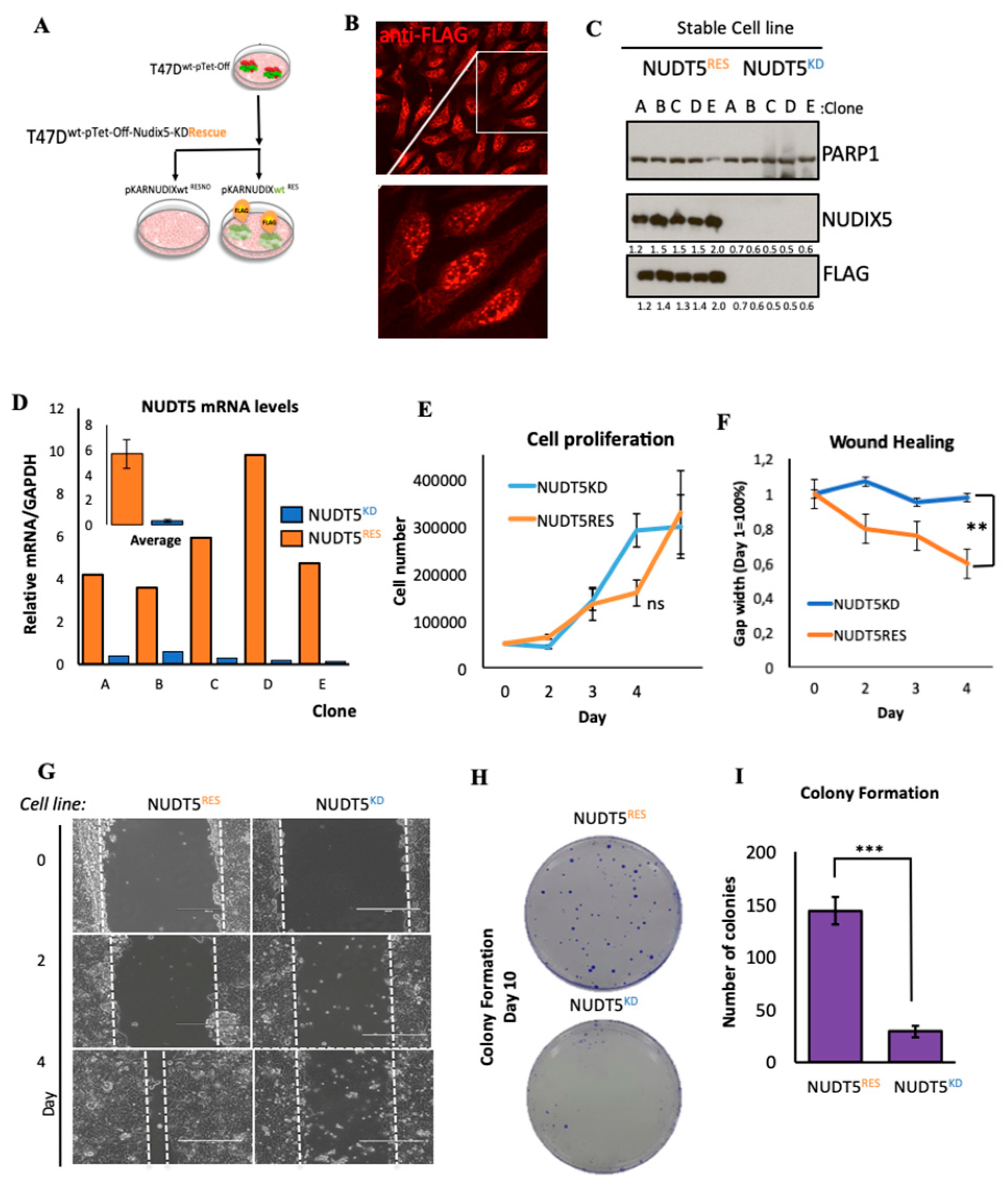
Cancers Free Full Text Expression Of Oncogenic Drivers In 3d Cell Culture Depends On Nuclear Atp Synthesis By Nudt5 Html
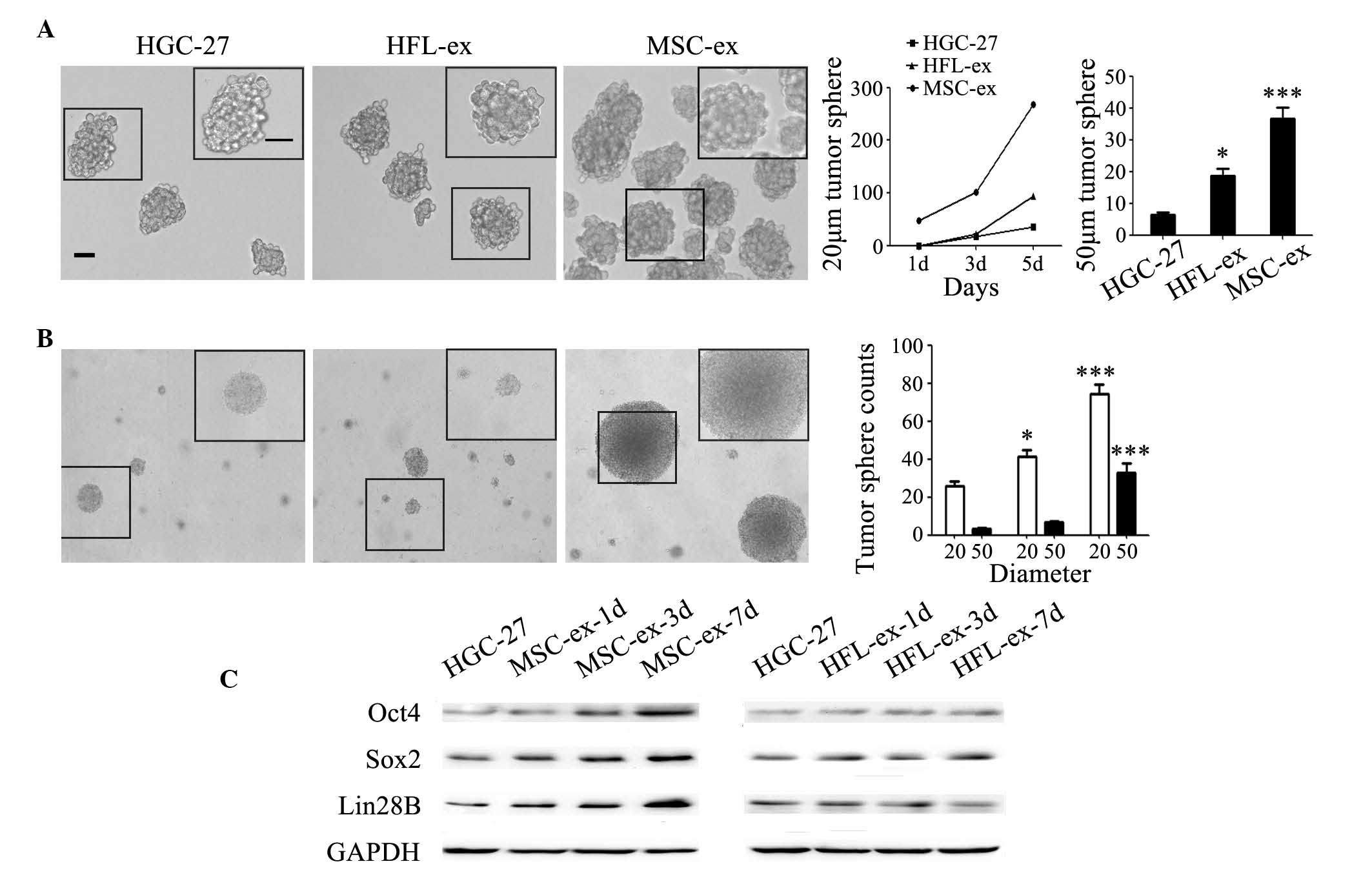
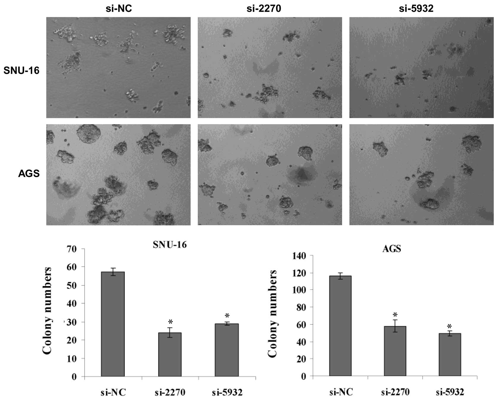
Exosomes Derived From Human Mesenchymal Stem Cells Promote Gastric Cancer Cell Growth And Migration Via The Activation Of The Akt Pathway
3d Matrix Based Cell Cultures Automated Analysis Of Tumor Cell Survival And Proliferation
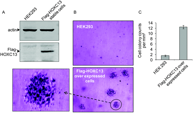
Antisense Oligonucleotide Mediated Knockdown Of Hoxc13 Affects Cell Growth And Induces Apoptosis In Tumor Cells And Over Expression Of Hoxc13 Induces Rsc Advances Rsc Publishing Doi 10 1039 C2ra206g
Biopply Com Assets References Cfa Abc Biopply Pdf

Proliferation And Invasion Plasticity In Tumor Cells Pnas

Effects Of Cdk9 Inhibition On Clonogenic Assay And 3d Culture A And Download Scientific Diagram
Jcm Free Full Text In Vitro Characterization Of Dental Pulp Stem Cells Cultured In Two Microsphere Forming Culture Plates Html

Glutathione Peroxidase 7 Gpx7 Suppresses Anchorage Independent Download Scientific Diagram

Preclinical Assessment Of The Bioactivity Of The Anticancer Coumarin Ot48 By Spheroids Colony Formation Assays And Zebrafish Xenografts Protocol
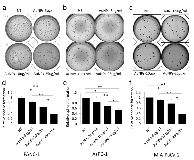
Gold Nanoparticles Sensitize Pancreatic Cancer Cells To Gemcitabine

Knockdown Of Cdh1 In Sw480 Cells Enhanced The Colony Formating Ability Download Scientific Diagram
P53 Is Involved In A Three Dimensional Architecture Mediated Decrease In Chemosensitivity In Colon Cancer

Foxc1 Promotes Colony Formation And Growth In 3d Matrigel A Foxc1 Download Scientific Diagram
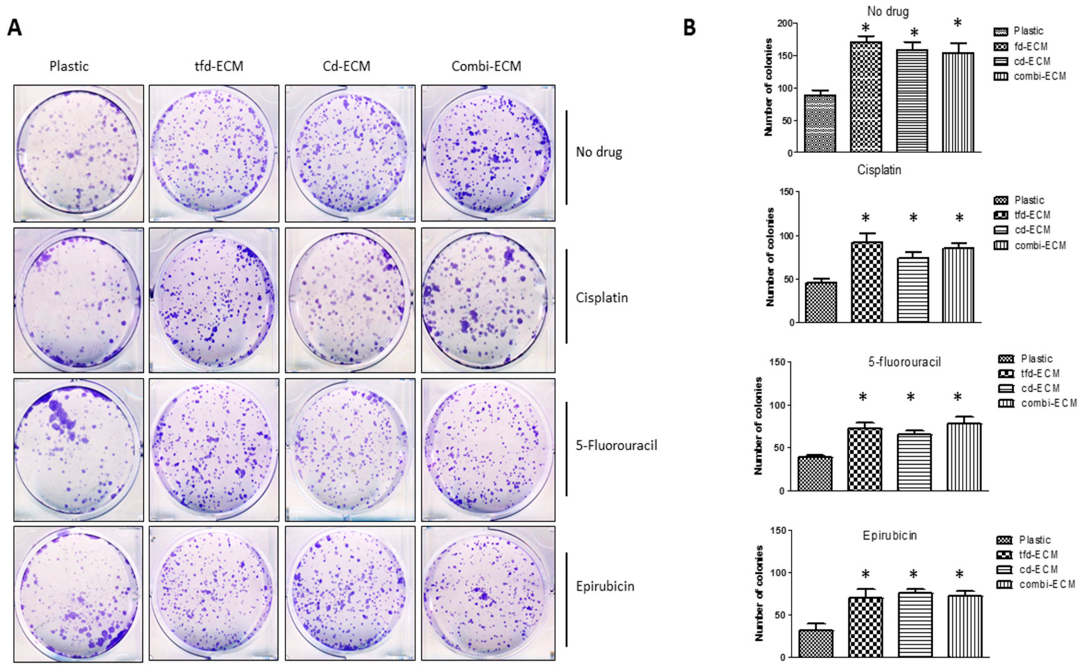
Ijms Free Full Text The Role Of Tumor Microenvironment In Chemoresistance 3d Extracellular Matrices As Accomplices Html
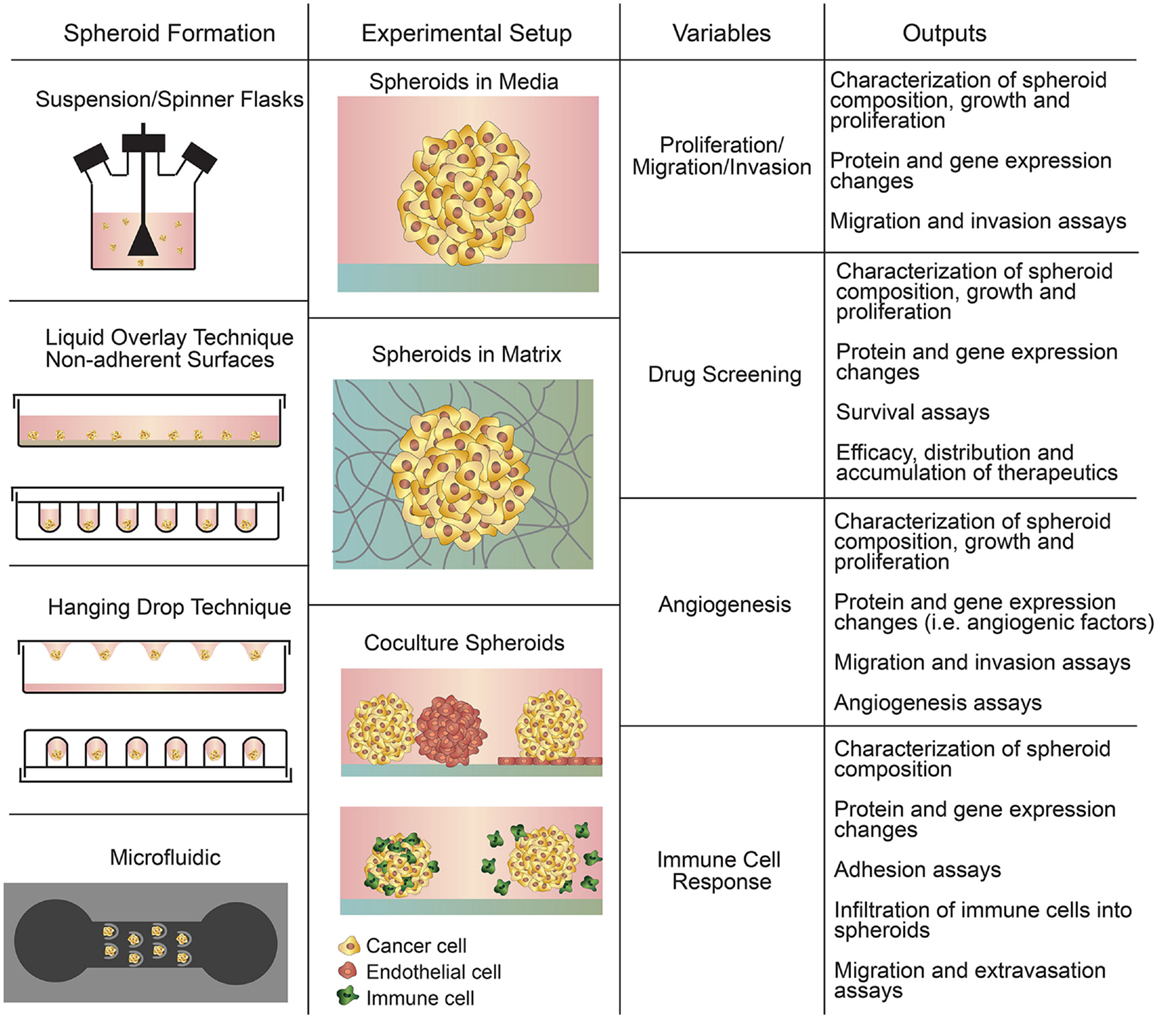
Frontiers In Vitro Tumor Models Advantages Disadvantages Variables And Selecting The Right Platform Bioengineering And Biotechnology
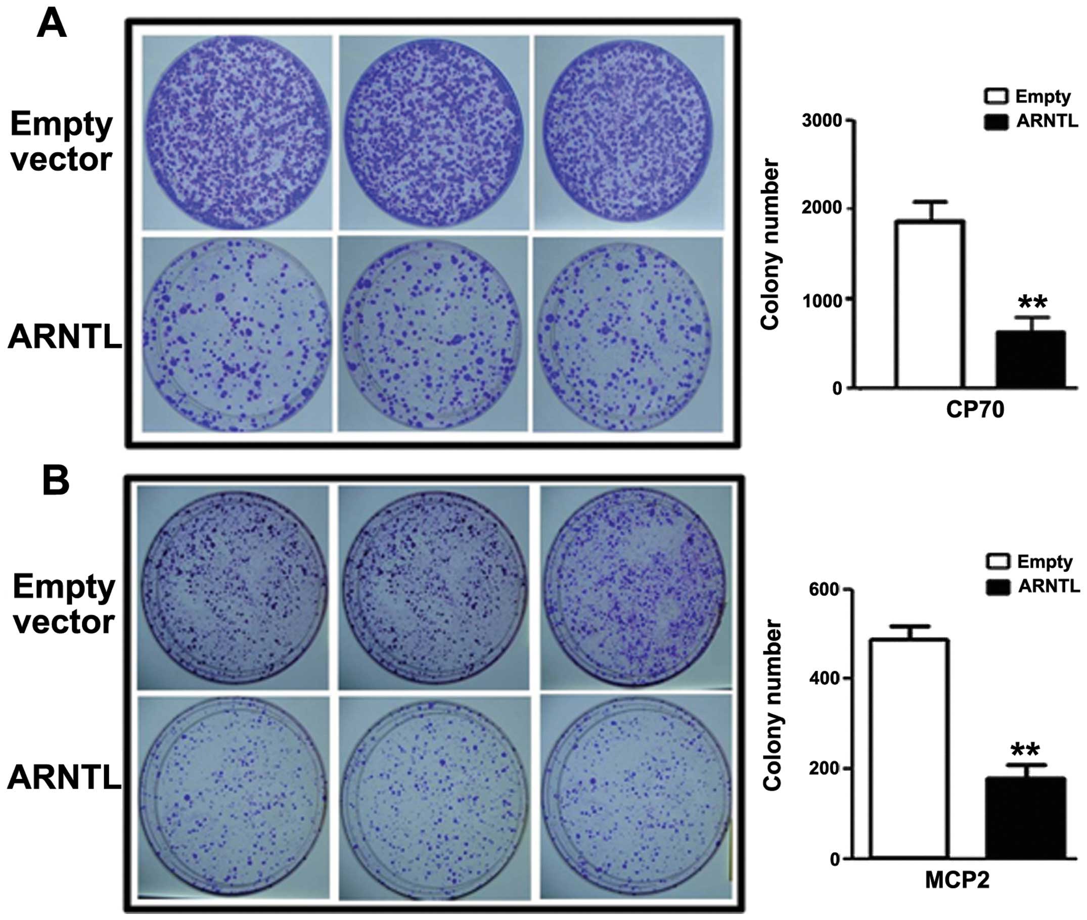
Epigenetic Silencing Of Arntl A Circadian Gene And Potential Tumor Suppressor In Ovarian Cancer
Journals Sagepub Com Doi Pdf 10 1177

Figure 2 From Strebloside Induced Cytotoxicity In Ovarian Cancer Cells Is Mediated Through Cardiac Glycoside Signaling Networks Semantic Scholar

Multiple Uses Of Basement Membrane Like Matrix Bme Matrigel In Vitro And In Vivo With Cancer Cells Benton 11 International Journal Of Cancer Wiley Online Library

Blockade Of Surface Bound Tgf B On Regulatory T Cells Abrogates Suppression Of Effector T Cell Function In The Tumor Microenvironment Science Signaling
Www 2bscientific Com Getmedia 48cf7444 f3 4bbe B1ca Df2df3e965 Colony Formation Brochure 14 Pdf
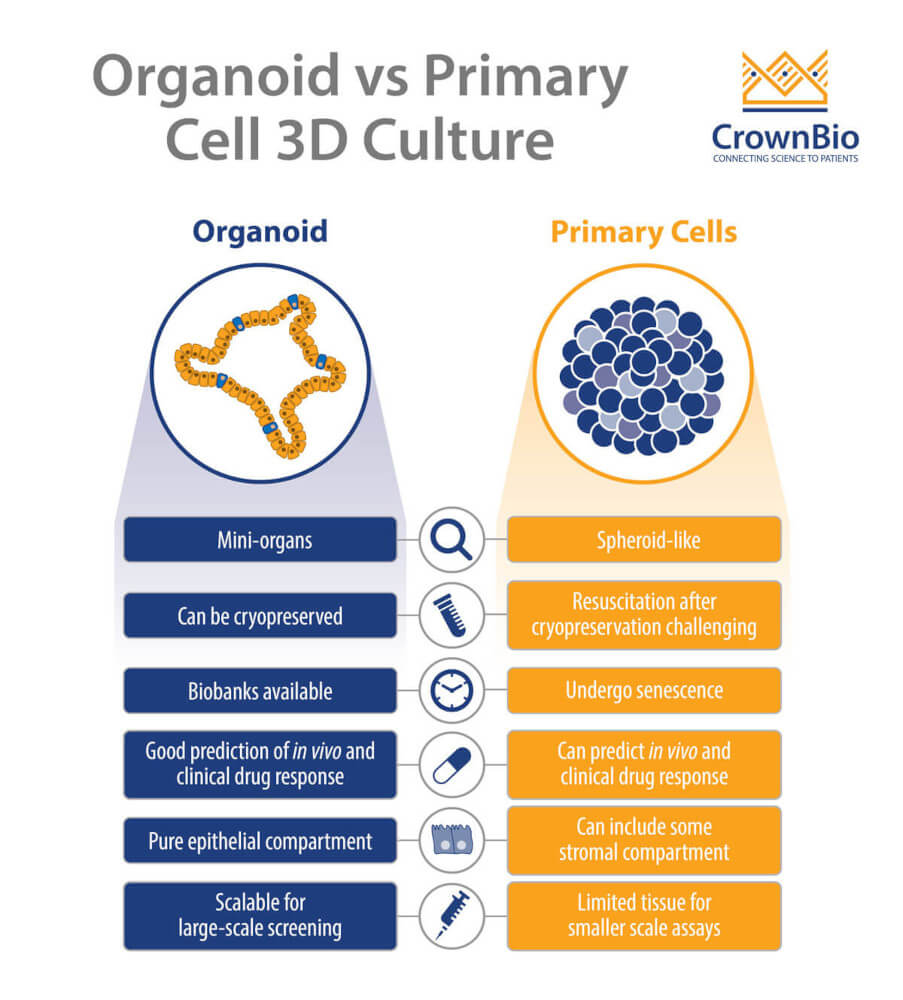
How Are Organoids Different From 3d Primary Cell Cultures
Knockdown Of Hmex 3a By Small Rna Interference Suppresses Cell Proliferation And Migration In Human Gastric Cancer Cells

Analysis Of Gsk J4 Post Treatment Effect By Colony Formation Assay Download Scientific Diagram

Oxford Optronix Gelcount
Www 2bscientific Com Getmedia 48cf7444 f3 4bbe B1ca Df2df3e965 Colony Formation Brochure 14 Pdf

The Human Colony Forming Cell Cfc Assay Using Methylcellulose Based Media R D Systems
Journals Sagepub Com Doi Pdf 10 1177

The Soft Agar Colony Formation Assay Protocol

Figure 1 From Automated Soft Agar Assay For The High Throughput Screening Of Anticancer Compounds Semantic Scholar
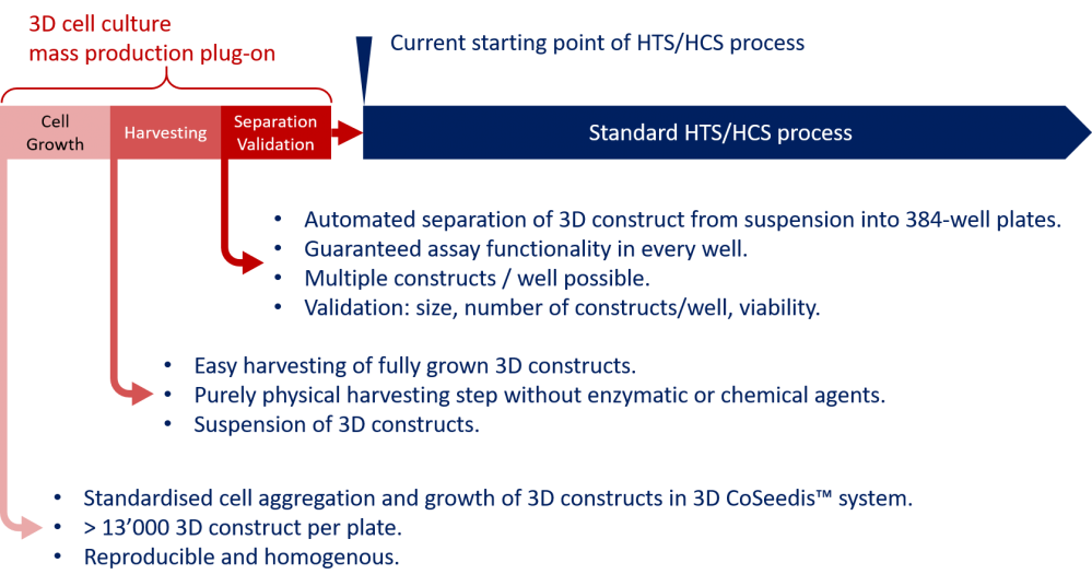
3d Coseedis System Bioke

Cells With Surface Expression Of Cd133highcd71low Are Enriched For Tripotent Colony Forming Progenitor Cells In The Adult Murine Pancreas Sciencedirect
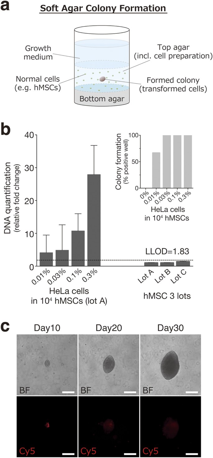
Ultra Sensitive Detection Of Tumorigenic Cellular Impurities In Human Cell Processed Therapeutic Products By Digital Analysis Of Soft Agar Colony Formation Scientific Reports

Proliferation And Invasion Plasticity In Tumor Cells Pnas

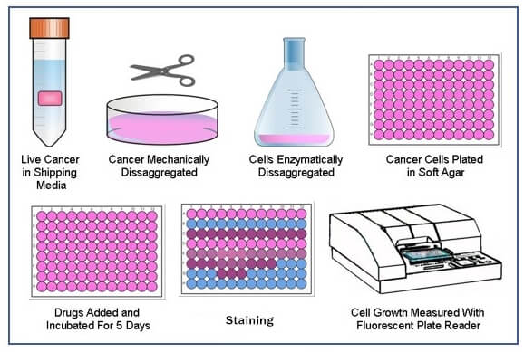
High Throughput Fluorescent Colony Formation Assay January 10

Improving The Metabolic Fidelity Of Cancer Models With A Physiological Cell Culture Medium Science Advances
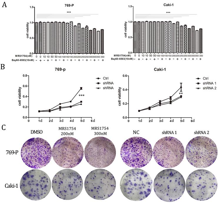
Blockade Of Adenosine b Receptor Reduces Tumor Growth And Migration In Renal Cell Carcinoma
Clonogenic Assay Creative Bioarray Creative Bioarray

A For 2d Or 3d Clonogenic Assays Single Cells Were Plated Onto Lrecm Download Scientific Diagram

A For 2d Or 3d Clonogenic Assays Single Cells Were Plated Onto Lrecm Download Scientific Diagram

An Hts Compatible 3d Colony Formation Assay To Identify Tumor Specific Chemotherapeutics Semantic Scholar
Inhibition Of Mir 96 Expression Reduces Cell Proliferation And Clonogenicity Of Hepg2 Hepatoma Cells
Cell Based Soft Agar Assays Reaction Biology
Biopply Com Assets References Cfa Abc Biopply Pdf
1
Targeting Of B1 Integrins Impairs Dna Repair For Radiosensitization Of Head And Neck Cancer Cells Oncogene

Growth Of Epithelial Organoids In A Defined Hydrogel Broguiere 18 Advanced Materials Wiley Online Library

3d Coseedis A New Approach To The Colony Forming Assay

Tumor Spheroid Formation Assay Sigma Aldrich
96 Well Cell Transformation Assays Standard Soft Agar

Tumor Spheroid Formation Assay Sigma Aldrich
Frontiers Sphere Formation Assay Three Dimensional In Vitro Culturing Of Prostate Cancer Stem Progenitor Sphere Forming Cells Oncology

3d Mammary Colony Forming Cell Assay Bio Protocol
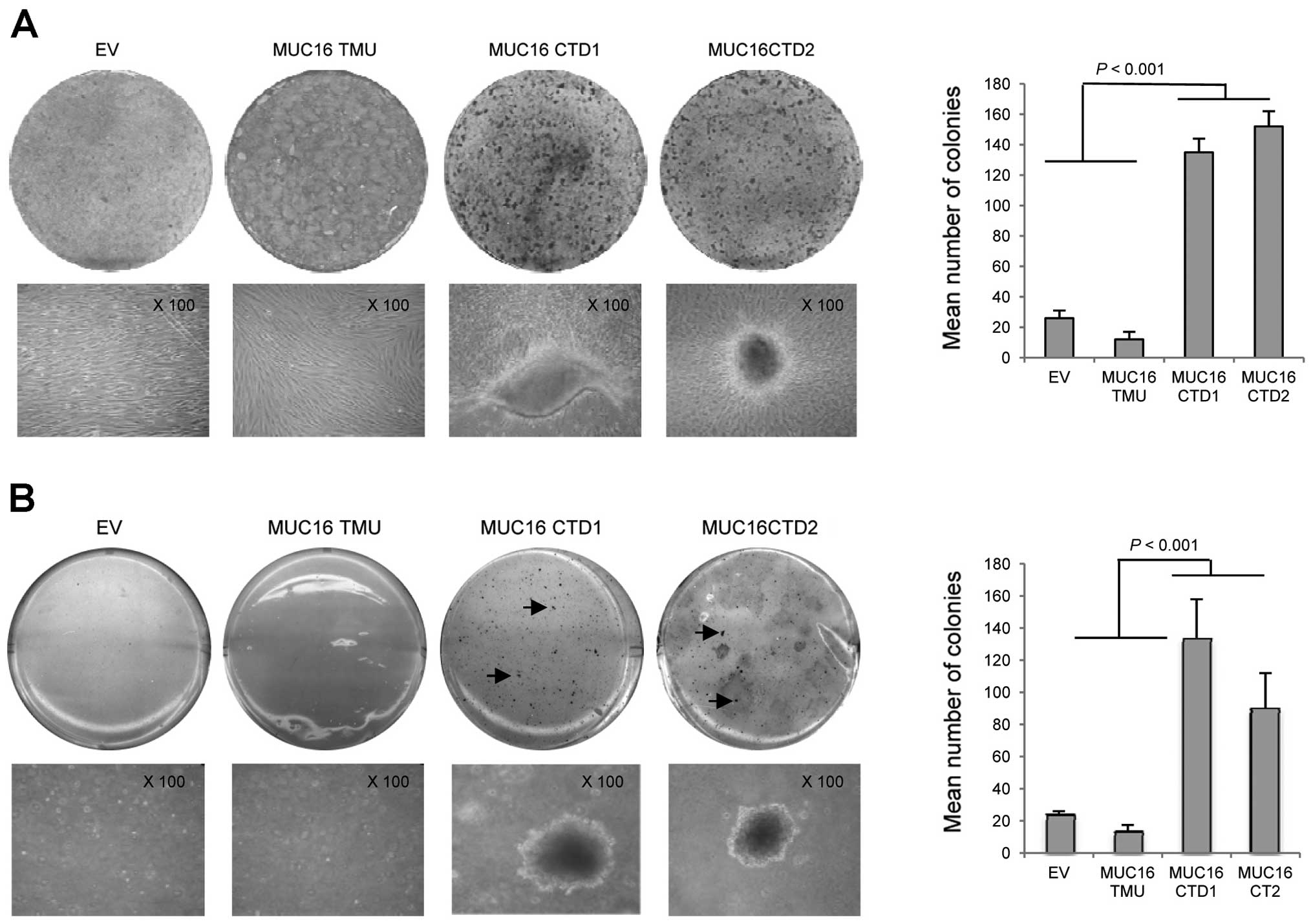
Transformation Of Nih3t3 Mouse Fibroblast Cells By Muc16 Mucin Ca125 Is Driven By Its Cytoplasmic Tail
Knockdown Of Setdb1 Inhibits Breast Cancer Progression By Mir 381 3p Related Regulation

Colony Forming Ability Of Three Glioma Cells Cultured Under 2d And 3d Download Scientific Diagram
2

Grb7 Inhibition Attenuates Tnbc Cell Motility Invasion And 3d Culture Download Scientific Diagram

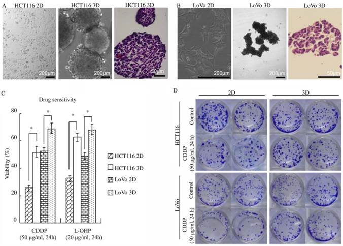
E Cadherin Knockdown In Hct116 Reduces Chemosensitivity Only In 3d Download Scientific Diagram

Tumor Spheroid Formation Assay Sigma Aldrich
Medicina Free Full Text Pleurotus Highking Mushroom Induces Apoptosis By Altering The Balance Of Proapoptotic And Antiapoptotic Genes In Breast Cancer Cells And Inhibits Tumor Sphere Formation Html

An Hts Compatible 3d Colony Formation Assay To Identify Tumor Specific Chemotherapeutics Semantic Scholar
In Vitro Analysis Of Breast Cancer Cell Line Tumourspheres And Primary Human Breast Epithelia Mammospheres Demonstrates Inter And Intrasphere Heterogeneity

Cancer Stem Cells From Colorectal Cancer Derived Cell Lines Pnas

Utilization Of The Soft Agar Colony Formation Assay To Identify Inhibitors Of Tumorigenicity In Breast Cancer Cells Protocol

Utilization Of The Soft Agar Colony Formation Assay To Identify Inhibitors Of Tumorigenicity In Breast Cancer Cells Protocol

An Hts Compatible 3d Colony Formation Assay To Identify Tumor Specific Chemotherapeutics Semantic Scholar

3d Mammary Colony Forming Cell Assay Bio Protocol
Www 2bscientific Com Getmedia 48cf7444 f3 4bbe B1ca Df2df3e965 Colony Formation Brochure 14 Pdf
Clonogenic Assay Creative Bioarray Creative Bioarray
Novel 3d Liquid Cell Culture Method For Anchorage Independent Cell Growth Cell Imaging And Automated Drug Screening Scientific Reports
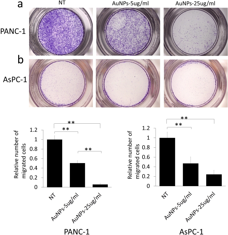
Gold Nanoparticles Sensitize Pancreatic Cancer Cells To Gemcitabine

High Throughput Fluorescent Colony Formation Assay January 10
Q Tbn And9gcrx2tyiisivvs32ze9286ip8lvxmyhc6rscr4ooytvrakmikmyo Usqp Cau
A New Dimension Of Cell Culture The Rise Of Spheroid Culture Systems Cell Culture Dish
Soft Agar Colony Formation Assay Creative Bioarray Creative Bioarray

Preclinical Assessment Of The Bioactivity Of The Anticancer Coumarin Ot48 By Spheroids Colony Formation Assays And Zebrafish Xenografts Protocol
Organotypic Culture Assays For Murine And Human Primary And Metastatic Site Tumors Nature Protocols
Plos Genetics Mir144 451 Expression Is Repressed By Runx1 During Megakaryopoiesis And Disturbed By Runx1 Eto
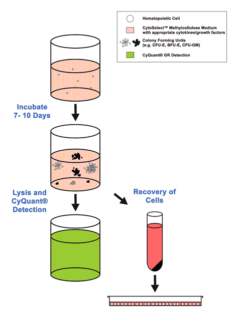
Hematopoietic Cfc Assays

Knockdown Of Cldn1 Inhibits Cell Growth In Soft Agar 3d But Not In Download Scientific Diagram

Establishment And Characterization Of Irradiated Hnscc Sublines A 3d Download Scientific Diagram

High Throughput Fluorescent Colony Formation Assay January 10

Oxford Optronix Gelcount
Exosomes Derived From Human Mesenchymal Stem Cells Promote Gastric Cancer Cell Growth And Migration Via The Activation Of The Akt Pathway
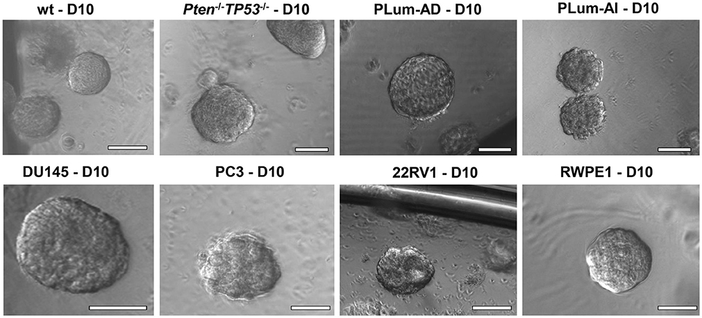
Frontiers Sphere Formation Assay Three Dimensional In Vitro Culturing Of Prostate Cancer Stem Progenitor Sphere Forming Cells Oncology

Bemer Electromagnetic Field Therapy Reduces Cancer Cell Radioresistance By Enhanced Ros Formation And Induced Dna Damage Abstract Europe Pmc

T47d A18 Pkca Cells Are Resistant To 4 Oht And Ral In 2d And 3d Cell Download Scientific Diagram
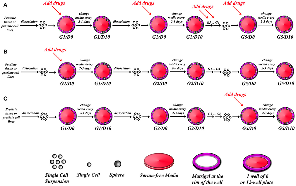
Frontiers Sphere Formation Assay Three Dimensional In Vitro Culturing Of Prostate Cancer Stem Progenitor Sphere Forming Cells Oncology
Potential Of Polycaprolactone Nanofiber Scaffold For Ex Vivo Expansion Of Cord Blood Derived Cd34 Hematopoietic Stem Cells


