Mrt T1 T2 Unterschied

3 Radiologische Diagnostik Des Multiplen Myeloms 3 3 Radiologisch Pathologische Korrelation
Mr Kontrastmittel Radiology Key
Opendata Uni Halle De Bitstream 1 Rdissertationen nach klinikenelektronische dissertationendigitalisierungdigitalisatedissertation Steffen Bauch Pdf

Magnetresonanztomographie Wikipedia
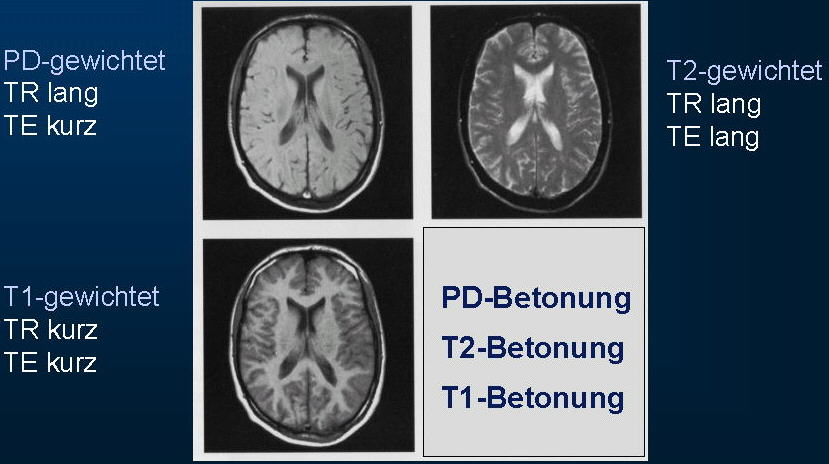
Bildgewichtung Und Kontrast
Das Bessere Bild Kommunikation Und Marketing Universitat Heidelberg
Das T2Verfahren ist eines der beiden Versandverfahren, mit der die grenzüberschreitende Beförderung von Waren innerhalb des Zollgebietes der Europäischen Union erfolgen kann Normalerweise fallen beim Warentransport innerhalb der EU keine Zölle und Einfuhrumsatzsteuern an Es bedarf insofern keiner besonderen Verfahrensregeln.
Mrt t1 t2 unterschied. In the preceding Q&A, T2 was defined as a time constant for the decay of transverse magnetization arising from natural interactions at the atomic or molecular levels used as a measurement of those processes contributing to the transverse decay of the MR signal that arise from natural interactions at the atomic and molecular levels within the tissue or substance of interest. "von T1 nach T2 wird das Licht angeschaltet" T1 Wasser dunkel,. T1, T2 or FLAIR) to highlight or suppress different types of tissue so that abnormalities can be detected Hyperintensity on a T2 sequence MRI basically means that the brain tissue in that.
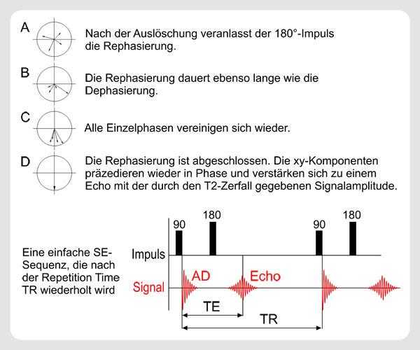
1GRE and SE can both provide T2* contrast 2GRE and SE use the same TE and TR to produce a T1weighted image 3SE is better for visualizing tissues with a very short T2 because of the refocusing pulses 4In GRE higher flip angles always produce brighter images Gradient Echoes & Flow. 26 MRTGeräteparameter Durch Veränderung der Geräteparameter (MRTSystemparameter), zB der Pulswiederholzeit (TR) oder der Echozeit (TE) kann man am MRT unterschiedliche Wichtungen einstellen T1gewichtete Bilder (T1w) T2gewichtete Bilder (T2w) Protonendichtegewichtete Bilder (PDGewichtung). Signal weighting (T1, T2, PD) and sequences parameters TR, TE;.
Definitions Scan of T1 MRI and T2 MRI T1 MRI, or T1weighted MRI, provides images with the contrast that is derived from the longitudinal time of relaxation of the explored soft tissue of the human organismThe shorter the relaxation time is, the brighter the resulting images T2 MRI, or T2weighted MRI, provides images with the contrast based on the T2, or transverse relaxation time of the. Merksprüche „T1 und T2 ist wie SchwarzWeißSehen von Flüssigkeiten“ und „ H 2 O ist in T 2 hyperintens (h ell)“!. In der T1Wichtung stellt sich Wasser hypointens dar, in der T2Wichtung ist es hyperintens!.
T2 bright/highintensity signal, usually greater than on T1, due to its high water content T1 C significant enhancement is seen due to high vascularity;. Both signal intensity ratios and electron microscopic features may be prognostic factors CDT celldense type MRT matrixrich type OS overall survival PFS progressionfree survival RT1, RT2, and REN ratios of tumortopons signal intensity in the T1 FLAIR sequence, T2 sequence, and enhanced T1 FLAIR sequence, respectively. T1, T2, and magnetization transfer (MT) measurements were performed in vitro at 3 T and 37°C on a variety of tissues mouse liver, muscle, and heart;.
Thickened trabeculae appear as low signal areas in both T1 and T2 images T1 highintensity signal due to its fat component;. CISS sequence uses a strong T2weighted 3D gradient echo technique which produces high resolution isotropic images Two consecutive runs of 3D balanced steadystate free precession with different excitation levels are performed internally and subsequently combined Image contrast in CISS is determined by the T2/T1 ratio of the tissue. Let Result1, Result2 be two variables, where Result1 is produced after running T1 and then T2, ie T1>T2;(serially) Result2 is produced after running T2 and then T1, ie T2>T1;(serially) Now suppose we interleave the actions of the two transaction, let us call it schedule S, now if the net result produced after running S is equivalent to.
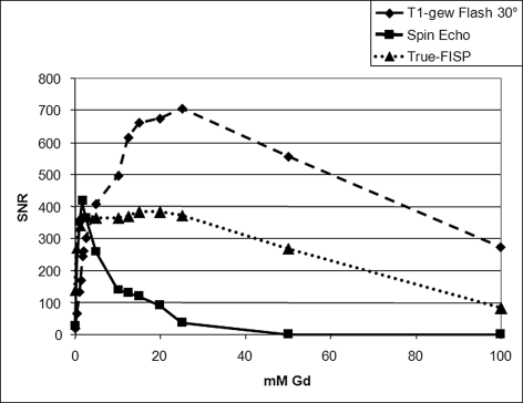
Bildgebende Verfahren Worin besteht der Unterschied in der Darstellung bei T1 und T2gewichteten MRTBilder bezogen auf Wasser und Fett?. The image contrast is a function of T1/T2 However, with a short TR and a short TE, the T1 portion remains constant The images are primarily T2weighted TrueFISP is very sensitive to the inhomogeneities in the magnetic field The images may contain interference stripes. The T1 and T2 values of 40 specimens from four femoral heads ranged from 386 to 1157 msec and from 54 to 139 msec, respectively.
What is the Difference between T1 & T2 “Customs Transit” Documents?. T 1,T 2 Relaxation and Magnetization Transfer in Tissue at 3T Greg J Stanisz,1,2* Ewa E Odrobina, 1Joseph Pun, Michael Escaravage,1 Simon J Graham, 1,2Michael J Bronskill, and R Mark Henkelman,1,2,3 T 1,T 2, and magnetization transfer (MT) measurements were performed in vitro at 3 T and 37°C on a variety of tissues mouse liver, muscle, and heart;. Learn about T1 vs T2 MRI scans with Pixorize's highyield visual mnemonics Part of our radiology playlist for medical school and the NBME shelf examsSubscr.
According to my knowledge , we use the difference between T1 and T2 in MRI to make contrast but I have no idea about The T2* time and what is the usage of that. "von T1 nach T2 wird das Licht angeschaltet" T1 Wasser dunkel,. Die MRT mit T1 und T2*Mapping zur Verifizierung einer Steatosis Hepatis ein Vergleich mit InPhase und OpposedPhaseSequenzen unterschied sich nicht signifikant von der medullaren und.
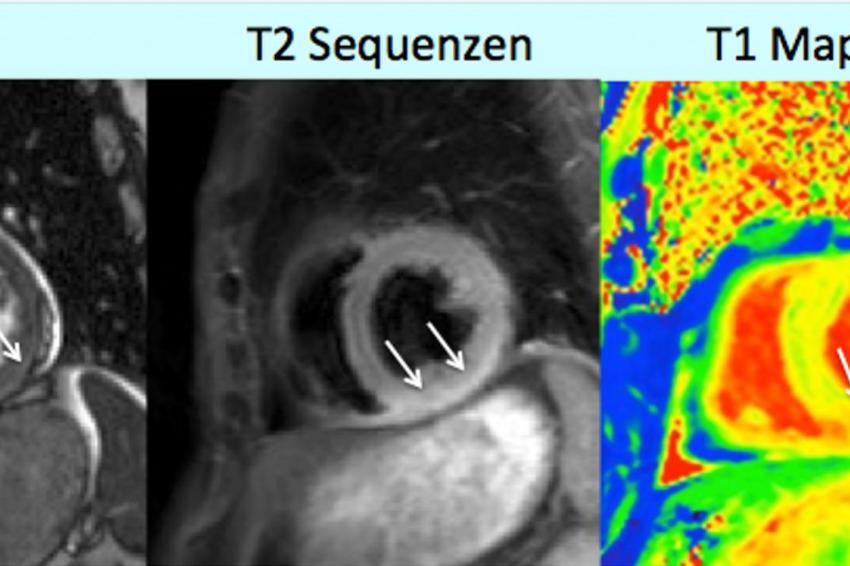
T1 relaxation is known as longitudinal relaxation and T2 is known as spinspin relaxation In the T1 type of relaxation, atomic nuclei come to thermal equilibrium in the magnetic field In the T2 process spins cause the irreversible loss of mechanization and cannot be recovered T1 MRI and T2 MRI produce different kinds of images. T1, T2 mapping and ECV can quantify diffuse, global myocardial pathologies Alterations of myocardial T1 and T2 relaxation times occur in various myocardial diseases (e g acute myocarditis) In the future mapping might act as a prognosticator or therapy monitoring tool Citation Format Roller FC, Harth S, Schneider C etal T1, T2 Map. A thorough explanation of these documents can be found in.
The image contrast is a function of T1/T2 However, with a short TR and a short TE, the T1 portion remains constant The images are primarily T2weighted TrueFISP is very sensitive to the inhomogeneities in the magnetic field The images may contain interference stripes. Rat spinal cord and kidney;. Therefore, 15 T and 3 T are correct Conversely, 15T and 3T are incorrect, despite the latter usages often being seen in medical media and some radiology reports.
Initially, the mural hematoma is isointense to the sternocleidomastoid muscle on T1WI and hypointense on T2 and TOF imaging At a mean delay of 5,8 days, mural hematoma may appear hyperintense on all sequences (Figs 7 and 8) Progressively, the signal drops on all pulse sequences 56 Only gold members can continue reading. Bildgebende Verfahren Worin besteht der Unterschied in der Darstellung bei T1 und T2gewichteten MRTBilder bezogen auf Wasser und Fett?. T1, T2 mapping and ECV can quantify diffuse, global myocardial pathologies Alterations of myocardial T1 and T2 relaxation times occur in various myocardial diseases (e g acute myocarditis) In the future mapping might act as a prognosticator or therapy monitoring tool Citation Format Roller FC, Harth S, Schneider C etal T1, T2 Map.
The FGATIR provides significantly better highresolution thin (1mm) slice visualization of DBS targets than does either standard 3T T1 or T2weighted imaging The FGATIR scans allowed for localization of the thalamus, striatum, GPe/GPi, Red Nucleus (RN), and Substantia Nigra (SNr) and displayed sharper delineation of these structures. Each MRI image consists of a T1 component and a T2 component (see also Relaxation section) It is possible to switch off most of one of either components, creating a T1 weighted or T2 weighted image respectively A special form is the proton density (PD) weighted image This sequence enables the visualization of the number of protons per volume. 26 MRTGeräteparameter Durch Veränderung der Geräteparameter (MRTSystemparameter), zB der Pulswiederholzeit (TR) oder der Echozeit (TE) kann man am MRT unterschiedliche Wichtungen einstellen T1gewichtete Bilder (T1w) T2gewichtete Bilder (T2w) Protonendichtegewichtete Bilder (PDGewichtung).
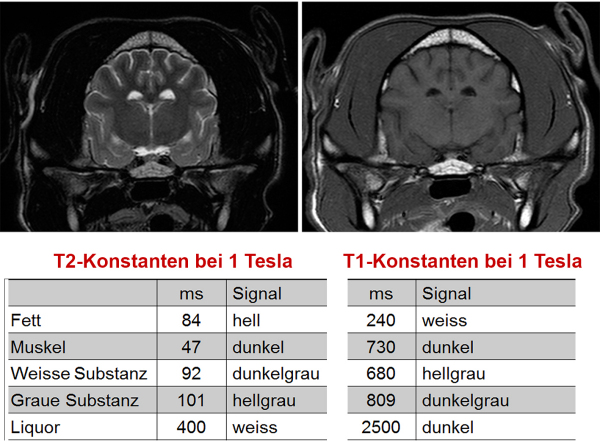
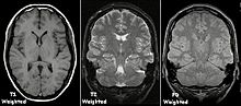
The T1 and T2 values of 40 specimens from four femoral heads ranged from 386 to 1157 msec and from 54 to 139 msec, respectively. Each MRI image consists of a T1 component and a T2 component (see also Relaxation section) It is possible to switch off most of one of either components, creating a T1 weighted or T2 weighted image respectively A special form is the proton density (PD) weighted image This sequence enables the visualization of the number of protons per volume. T1 und T2 sind entscheidend für den Bildkontrast Bei einer T1Gewichtung gibt es andere Bilder als bei einer T2Gewichtung (bei B0=1,5T) T1 (ms) 870 490 650 780 260 >4000 0 T2 (ms) 47 43 58 67 84 >00 79 Skelettmuskel Leber Niere Milz Fett Liquor Lunge Abb 7 T1 und T2Relaxation Abb 8 T1/T2 in verschiedenen Medien T1Relaxation.
The simple answer is that T1, T2, and FLAIR are different mathematical formulas for throwing those magnets around They spin in different directions, depending upon which formula you use For the health page, look at the top of the screen, on the right hand side How An MRI Works. T1MRT und T2MRT erzeugen unterschiedliche Arten von Bildern Schauen wir uns diese Unterschiede genauer an Definitionen T1 MRTDie T1gewichtete MRT liefert Bilder mit dem Kontrast, der sich aus der Längszeit der Relaxation des untersuchten Weichgewebes des menschlichen Organismus ergibt Je kürzer die Relaxationszeit ist, desto heller sind die resultierenden Bilder. I've actually written a health page on this one!.
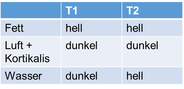
The two basic types of MRI images are T1weighted and T2weighted images, often referred to as T1 and T2 images On T1 images FAT is white On T2 images both FAT and WATER are white. The two basic types of MRI images are T1weighted and T2weighted images, often referred to as T1 and T2 images On T1 images FAT is white On T2 images both FAT and WATER are white. Radiologie 5 MRT Unterschiede zw T1 und T2 Wichtung CAVE egal ob T1 / T2, Fett ist immer hyperintens (= signalreich = hell) entscheidend ist die Darstellung von Flüssigkeit (T2 = ZWEIss).
MRI of the Cervical Spine, sagittal T2weighted image 1, Lateral mass of C1 (atlas) 2, Posterior arch of C1 3, Vertebral foramen with cerebrospinal fluid 4, Spinous process of l'axis Image 12 MRI of the Cervical Spine, sagittal T2weighted image 1, Vertebral foramen Cerebrospinal fluid. Basics of tissue contrast in MRI;. T1 relaxation is known as longitudinal relaxation and T2 is known as spinspin relaxation In the T1 type of relaxation, atomic nuclei come to thermal equilibrium in the magnetic field In the T2 process spins cause the irreversible loss of mechanization and cannot be recovered T1 MRI and T2 MRI produce different kinds of images.
Each MRI image consists of a T1 component and a T2 component (see also Relaxation section) It is possible to switch off most of one of either components, creating a T1 weighted or T2 weighted image respectively A special form is the proton density (PD) weighted image This sequence enables the visualization of the number of protons per volume. For example, the three properties, T1, T2, and B1, can lead to a Dictionary with over 700,000 entries A pattern matching process compares the fingerprints with the Dictionary When there is a match, the properties of this fingerprint are assigned to a map This process is repeated sequentially until all fingerprints have a corresponding. Bovine optic nerve, c.
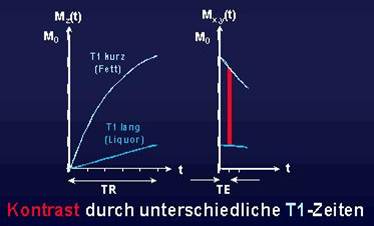
1 Definition Als T1Gewichtung bezeichnet man eine Kontrastdarstellung von MRTBildern, bei der die Repetitionszeit (TR) und die Echozeit (TE) so gewählt werden, dass die untersuchten Gewebe vor allem durch ihre T1Relaxationszeit, und weniger ihre T2Relaxationszeit differenziert werden siehe auch T2Gewichtung 2 Physikalische Grundlagen Bei jeder Bildakquisition wird die gewählte. T1 T2 PD Image Water signal Water has a long T1 T1WI uses a short TR so the signal from water is still low, therefore, water appears dark T2WI uses a long TE so the signal from water is high, therefore, water appears bright A long TR results in a high water signal, but a short TE means that this is less than the signal of a T2 scan. Alternative Methoden Magnetresonanzspektroskopie Abkürzungen MRSpektroskopie, MRS;.
T1 und T2 sind entscheidend für den Bildkontrast Bei einer T1Gewichtung gibt es andere Bilder als bei einer T2Gewichtung (bei B0=1,5T) T1 (ms) 870 490 650 780 260 >4000 0 T2 (ms) 47 43 58 67 84 >00 79 Skelettmuskel Leber Niere Milz Fett Liquor Lunge Abb 7 T1 und T2Relaxation Abb 8 T1/T2 in verschiedenen Medien T1Relaxation. Learn about T1 vs T2 MRI scans with Pixorize's highyield visual mnemonics Part of our radiology playlist for medical school and the NBME shelf examsSubscr. For each patient, axial tSWI and T2* sequences and conventional MR imaging sequences, such as T1, T2, and FLAIR, were performed tSWI was reconstructed to TSWI, by performing a minimumintensityprojection, by using postprocessing software in the PACS system, making it possible to adjust section thickness and setting it to the same section.
T1weighted inphase (a) and opposedphase (b) MR images show the lesion (arrow), which demonstrates homogeneous chemical shift signal intensity loss (arrow on b) on the opposedphase image The demographics of the patient, her history, and the absence of chronic liver disease allowed a confident diagnosis of HNF1α–mutated hepatocellular. In one study, Hanna et al compared MRI scans using T1weighted, T2weighted, STIR, and contrastenhanced T1weighted sequences with histologic specimens at 21 sites, 7 of which contained tumor and 14 of which were tumorfree For all of the tumorpositive sites, the MRI scans revealed abnormalities. Findings The T1 value positively correlated with the yield stress (σ y) and collapsed stress (σ c)The T2 value did not correlate with the yield stress, but it correlated with the collapsed stress and strength reduction ratio (σ c /σ y), which reflects the progressive refracture riskPartial correlation coefficient analyses, after adjusting for the bone mineral density, showed a.
Die Magnetresonanztomographie, abgekürzt MRT oder MR (als Tomographie von altgriechisch τομή tome, deutsch ‚Schnitt‘ und γράφειν graphein, deutsch ‚schreiben‘), ist ein bildgebendes Verfahren, das vor allem in der medizinischen Diagnostik zur Darstellung von Struktur und Funktion der Gewebe und Organe im Körper eingesetzt wird Es basiert physikalisch auf den Prinzipien. The average rate at which molecules tumble (and therefore T1 and T2 time) is related to the molecular size Small molecules (eg water/CSF) have a broad distribution of motional frequencies with poor matching with the Larmor frequency and therefore have long T1 values Medium sized molecules (eg lipids) have a narrower distribution of tumbling rates matched to typical resonant frequencies. Es ist oft schwer, den Unterschied zwischen einer Zyste und einem Tumor zu erkennen selbst für Ärzte Es gibt zwar ein paar Dinge, nach denen Sie suchen können, um festzustellen, ob ein Knoten eher eine Zyste oder ein Tumor ist Es empfiehlt sich jedoch, einen Termin mit Ihrem Arzt zu vereinbaren.
Radiologie 5 MRT Unterschiede zw T1 und T2 Wichtung CAVE egal ob T1 / T2, Fett ist immer hyperintens (= signalreich = hell) entscheidend ist die Darstellung von Flüssigkeit (T2 = ZWEIss). Terminology It is important to emphasize that in common with standard scientific unit notation, a space must always be inserted between the quantity and the unit symbol;. Rat spinal cord and kidney;.
T2*weighted imaging is built from the basic physics of magnetic resonance imaging where there is spin–spin relaxation, that is, the transverse component of the magnetization vector exponentially decays towards its equilibrium value It is characterized by the spin–spin relaxation time, known as T 2In an idealized system, all nuclei in a given chemical environment, in a magnetic field. The average rate at which molecules tumble (and therefore T1 and T2 time) is related to the molecular size Small molecules (eg water/CSF) have a broad distribution of motional frequencies with poor matching with the Larmor frequency and therefore have long T1 values Medium sized molecules (eg lipids) have a narrower distribution of tumbling rates matched to typical resonant frequencies. When transporting goods between countries, borders are crossed To ensure a smooth transit of goods all over the world, certain documents are needed BC Duty Free provides you with all the information you need!.
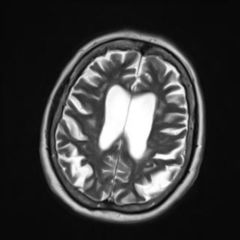
MRI of the Cervical Spine, sagittal T2weighted image 1, Lateral mass of C1 (atlas) 2, Posterior arch of C1 3, Vertebral foramen with cerebrospinal fluid 4, Spinous process of l'axis Image 12 MRI of the Cervical Spine, sagittal T2weighted image 1, Vertebral foramen Cerebrospinal fluid. Definition Verfahren, das den Spin von unterschiedlichen Protonen in einem definierten. A noncontrast enhanced, T2 weighted brain MRI using at least a 15 Tesla scanner and a noncontrast enhanced 3D volumetric T1weighted brain MRI will be performed at baseline for all PPMI subjects Therefore, it is required that the radiology center (or person identified as responsible) transmit the MRIs to the imaging core lab.
Pedirad

Magnetresonanzangiographie Wikiwand
Http Dbm Neuro Uni Jena De Pdf Files Gaser Morphometrie Pdf
Bildgebende Verfahren Rontgen Ct Und Mrt Kenhub
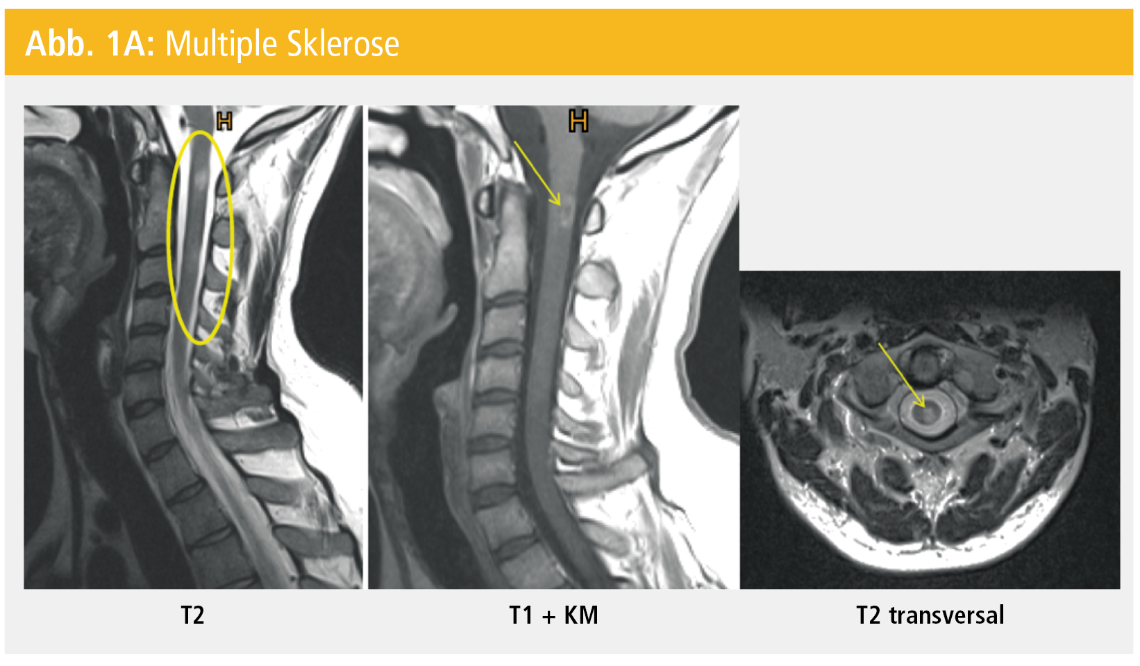
Neuroradiologie Zerebraler Gliome Bei Erwachsenen Rosenfluh Ch
Mrt Des Kopfes
Freidok Uni Freiburg De Fedora Objects Freidok 3315 Datastreams File1 Content
Opus Bibliothek Uni Wuerzburg De Files Evangelista Laura Dwi Mrt Pdf

Leberherde Im Mri
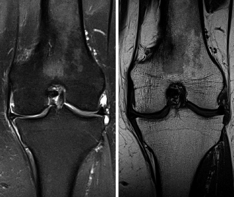
Wichtungen Und Sequenzen Der Magnetresonanztomographie In Der Traumatologie Springerlink
Strukturelle Bildgebung Des Gehirns

3 Radiologische Diagnostik Des Multiplen Myeloms 3 3 Radiologisch Pathologische Korrelation
Bildgewichtung Und Kontrast
Http Www Physik Uni Regensburg De Studium Medphys Nitz Mrt Hndout Ss18 V11 Red Pdf
Space Cube Vista Questions And Answers In Mri
Messsequenzen Fur Die Mr Bildgebung

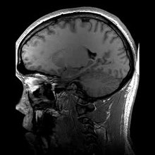
Magnetresonanztomographie Wikipedia

Kernspintomographie
Www Greenscan Imaging De App Download Roefo 3269de Pdf

Bildgebende Verfahren Faszination Mrt Klinik Via Medici

T2 Vs T2 Relaxation Time Questions And Answers In Mri
Bildgebende Verfahren Faszination Mrt Klinik Via Medici
Mrt Unterschiede

Bildgebende Verfahren Faszination Mrt Klinik Via Medici

Die Gliome Des Erwachsenen

Die Genauigkeit Der Magnetresonanztomographie Zur Vorhersage Eines Wanduberschreitenden Wachstums Des Rektalen Karzinoms Im Vergleich Zur Histologie Pdf Kostenfreier Download

Leberherde Im Mri
Q Tbn And9gctxqudh6wxnxxfxyayqqbg Rjb9fwue5cgpgdbexgsvntxpa Jl Usqp Cau
Mediziner Orthopadie Sind Gefragt Was Konnte Das Auf Dem Mrt Bild Sein Medizin Wirbelsaule Erkrankung
Neue Methoden Fur Neuro Mrt Philips Healthcare
Dgk Org Daten Radunski T1 T2 Mapping Text Pdf
Warum Mrt Springerlink
Myokarditis Mrt Hat Hochste Sensitivitat Und Spezifitat

Produktbroschure The Fine Art Of Liver Imaging Pdf Kostenfreier Download
Was Ist Eine Tr Mrt Repetico
Stellenwert Der Magnetresonanztomografie Beim Staging Des Harnblasenkarzinoms Medmedia
Http Bilder Buecher De Zusatz 819 Lese 1 Pdf
Hamangiome Des Knochens Springerlink
Core Ac Uk Download Pdf Pdf
Mediatum Ub Tum De Doc Pdf
Radiosurfvet

3 Radiologische Diagnostik Des Multiplen Myeloms 3 3 Radiologisch Pathologische Korrelation
Radiologie Der Gonarthrose Konventionelle Rontgendiagnostik Und Schnittbildgebung Asu
Dgn Org Wp Content Uploads 12 12 Ll 17 12 Vaskulre Demenzen Pdf
Kardiale Mrt Goldstandard Bei Fokalen Narben Management Krankenhaus

File Tektales Lipom 77jm Mrt Verschiedene Wichtungen Und Ebenen 001 Jpg Wikimedia Commons

Neuroradiologie Zerebraler Gliome Bei Erwachsenen Rosenfluh Ch
T1 Gewichtung Doccheck Flexikon
Http Www Imp Uni Erlangen De Lehre Mr imaging Pdf
Www Thieme Connect De Products Ebooks Pdf 10 1055 B 0033 773 Pdf

Magnetresonanztomographie Wikipedia
Mr Kontrastmittel Radiology Key
Docserv Uni Duesseldorf De Servlets Derivateservlet Derivate 2757 757 Pdf

Abb 8 A D 3 Die T1 Und T2 Gewichteten Mrt Bei Diagnosestellung A B Download Scientific Diagram
Www Ukgm De Ugm 2 Deu Umr Rdi Teaser Grundlagen Der Magnetresonanztomographie Mrt 13 Pdf
Www Studocu Com De Document Universitaet Bremen Klinische Neuropsychologie Zusammenfassungen Strukturelle Bildgebung Zusammenfassung View
Pedirad
Www Baltic Imaging Center De Images 267 264 Spm Kurs 12 Physik Pdf

Fat Suppression Methods Questions And Answers In Mri
D Nb Info 34
Q Tbn And9gcqcpmrx5ueluseruqmc47ivoyq Hyagbwkzzhqukymosfk7lkf5 Usqp Cau

Relaxationsprozesse In Der Nmr Spektroskopie Chemgapedia
T1 Flair Questions And Answers In Mri
Der Stellenwert Von Ultraschall Ct Und Mrt Ogpb
Magnetresonanztomographie Wikipedia

Wie Funktioniert Eigentlich T1 Und T2 Wichtung Im Mrt Youtube
Hamangiome Des Knochens Springerlink

Radiologie
Bildgebende Diagnostik Von Knochenmetastasen

Grundlagen Und Unterscheidungsmerkmale Zu Wichtigen Mrt Sequenzen Youtube
Radiosurfvet

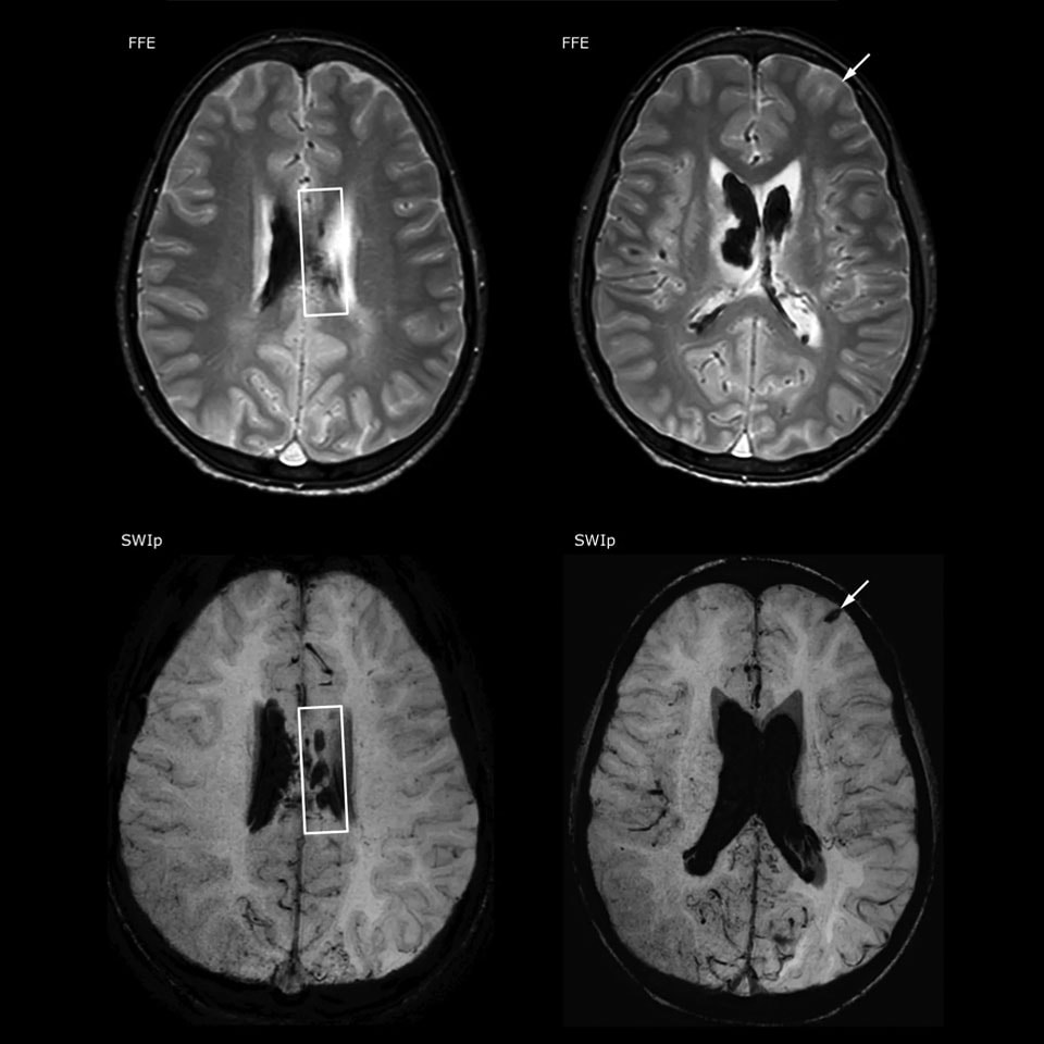
Klinische Anwendung Der Swi Sequenz In Der Neuroradiologie Pdf Kostenfreier Download
Compgefa Magnetresonanztomographie

Magnetresonanzangiographie Wikiwand
Www Ukgm De Ugm 2 Deu Umr Rdi Teaser Grundlagen Der Magnetresonanztomographie Mrt 13 Pdf
%20-%20Presented%20by%20PostDICOM.jpg)
Hier Ist Alles Was Sie Uber Ct Und Mrt Imaging Wissen Mussen Postdicom
Www Thieme Connect De Products Ebooks Pdf 10 1055 B 0035 Pdf

Inversion Recovery Sequenz Doccheck Flexikon
Mediatum Ub Tum De Doc Pdf
Magnetresonanztomographie Mrt Dein Online Radiologe
.jpg)
Bildgebende Verfahren Faszination Mrt Klinik Via Medici

Kontrastmittel Beim Mrt Der Multiplen Sklerose Medizin Therapie Multiple Sklerose News Amsel E V
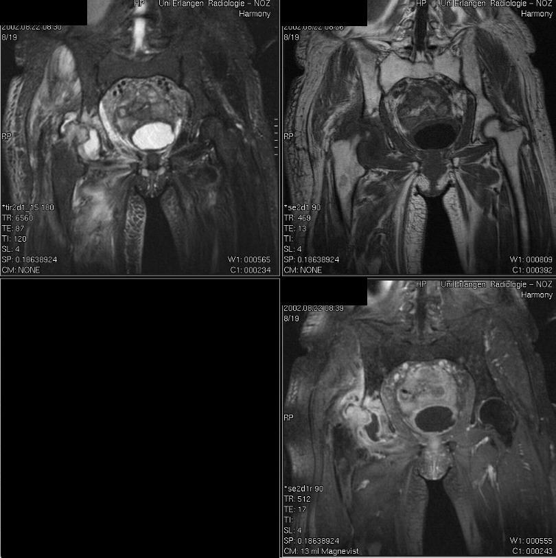
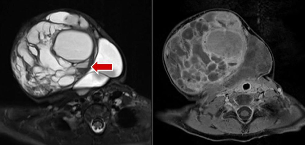
Abb 5a C 9 Mrt Bei On Beidseits Und Stiller Hufte Rechts A Download Scientific Diagram
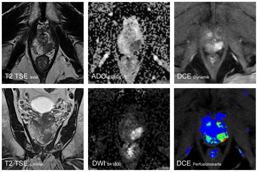
Multiparametrische Mrt Verbessert Die Diagnostik Des Prostatakarzinoms
Www Ukgm De Ugm 2 Deu Umr Rdi Teaser Grundlagen Der Magnetresonanztomographie Mrt 13 Pdf
Triple Inversion Recovery Cardiac Mri Questions And Answers In Mri

Abb 8 A D 3 Die T1 Und T2 Gewichteten Mrt Bei Diagnosestellung A B Download Scientific Diagram
Www Researchgate Net Profile Heike Daldrup Link Publication Mr Diagnostik Von Knochenmarkerkrankungen Links 57b9fae14f440bd8df3 Mr Diagnostik Von Knochenmarkerkrankungen Pdf
Magnetresonanztomographie Mrt Dein Online Radiologe
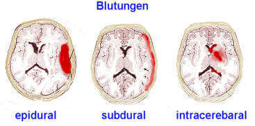
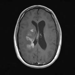
Blutungen Intracerebrale
Mrt
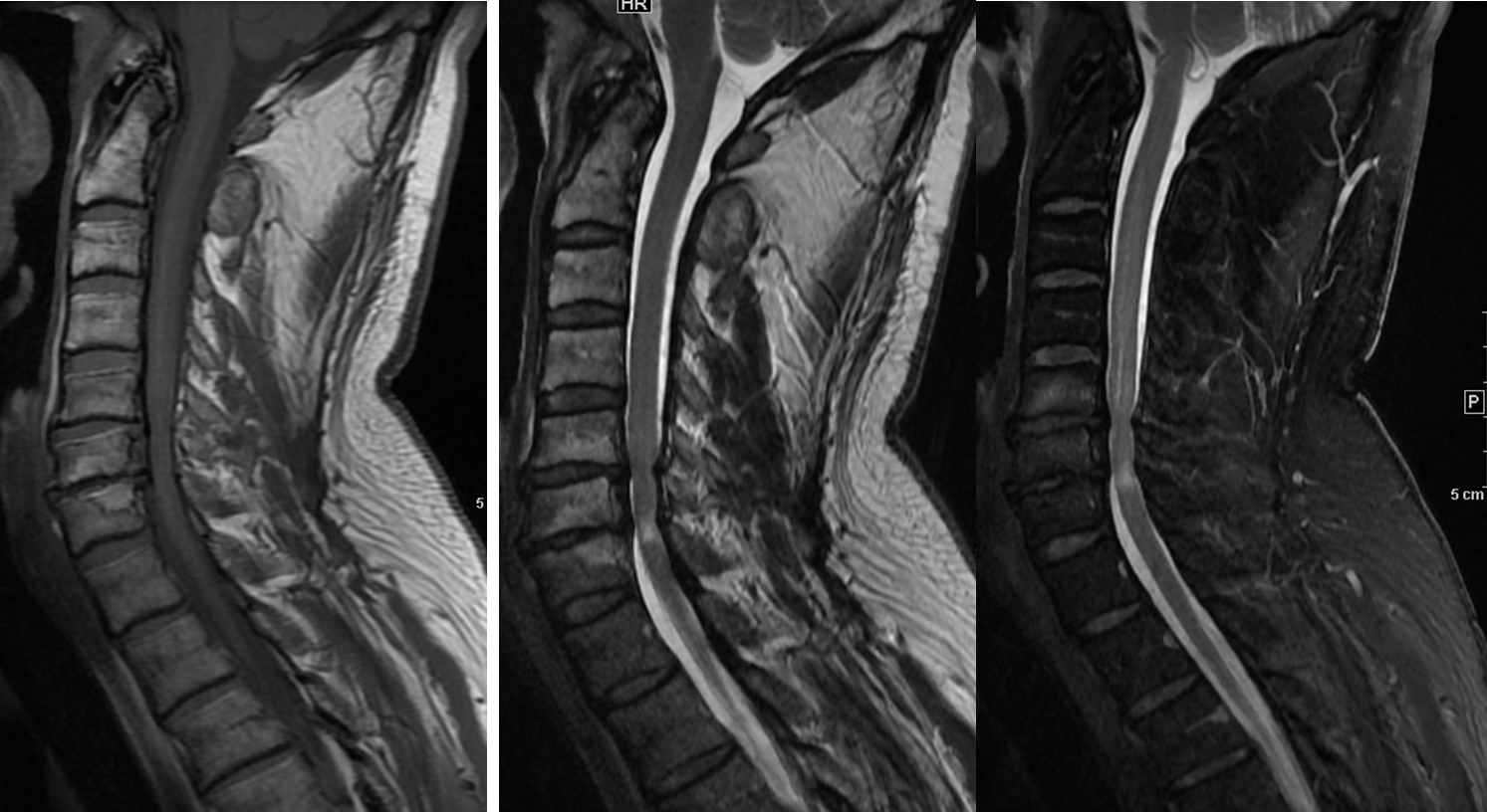
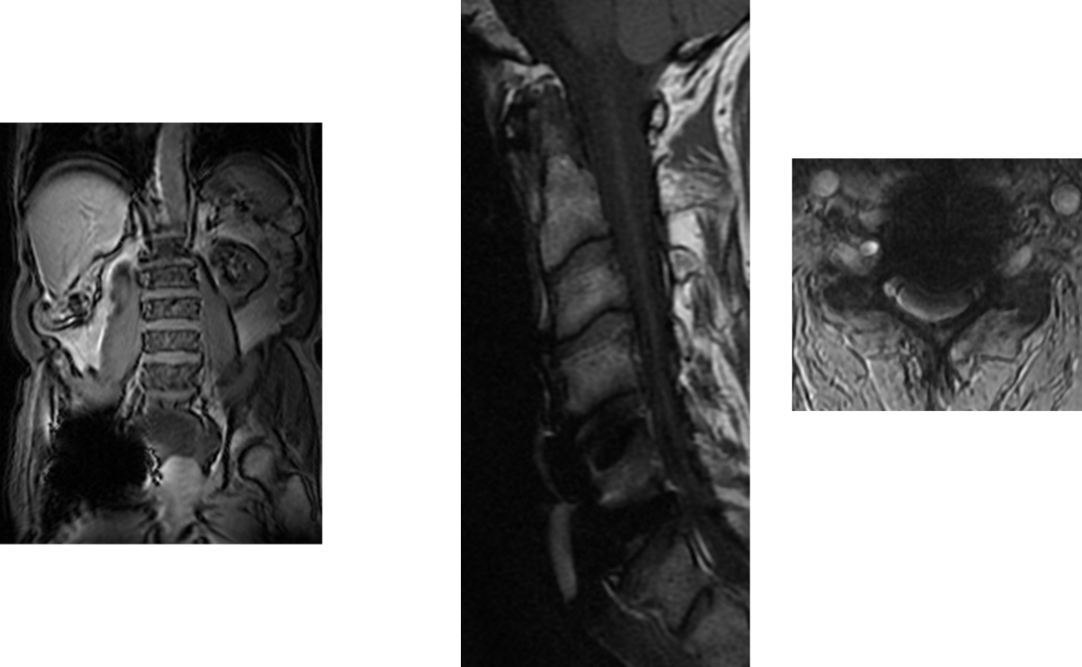
Mrt Diagnostik Von Ruckenmarkserkrankungen Medmedia
Magnetresonanztomographie Mrt Dein Online Radiologe
Erstellung Von Quantitativen T1 T2 Und Rheinahrcampus
T2 Gewichtung Doccheck Flexikon
Magnetresonanztomographie Wikipedia


